• 文献检索
  • 文档翻译
  • 深度研究
  • 学术资讯
  • Suppr Zotero 插件Zotero 插件
  • 邀请有礼
  • 套餐&价格
  • 历史记录
应用&插件
Suppr Zotero 插件Zotero 插件浏览器插件Mac 客户端Windows 客户端微信小程序
定价
高级版会员购买积分包购买API积分包
服务
文献检索文档翻译深度研究API 文档MCP 服务
关于我们
关于 Suppr公司介绍联系我们用户协议隐私条款
关注我们

Suppr 超能文献

核心技术专利:CN118964589B侵权必究
粤ICP备2023148730 号-1Suppr @ 2026

文献检索

告别复杂PubMed语法,用中文像聊天一样搜索,搜遍4000万医学文献。AI智能推荐,让科研检索更轻松。

立即免费搜索

文件翻译

保留排版,准确专业,支持PDF/Word/PPT等文件格式,支持 12+语言互译。

免费翻译文档

深度研究

AI帮你快速写综述,25分钟生成高质量综述,智能提取关键信息,辅助科研写作。

立即免费体验

OCT 立方体中与年龄相关的黄斑变性液体积聚的全自动 3D 直观可视化。

Fully-Automatic 3D Intuitive Visualization of Age-Related Macular Degeneration Fluid Accumulations in OCT Cubes.

机构信息

Grupo VARPA, Instituto de investigación Biomédica de A Coruña (INIBIC), Xubias de Arriba, 84, A Coruña, 15006, Spain.

Centro de investigación CITIC, Universidade da Coruña, Campus de Elviña, s/n, A Coruña, 15071, Spain.

出版信息

J Digit Imaging. 2022 Oct;35(5):1271-1282. doi: 10.1007/s10278-022-00643-6. Epub 2022 May 5.

DOI:10.1007/s10278-022-00643-6
PMID:35513586
原文链接:https://pmc.ncbi.nlm.nih.gov/articles/PMC9582110/
Abstract

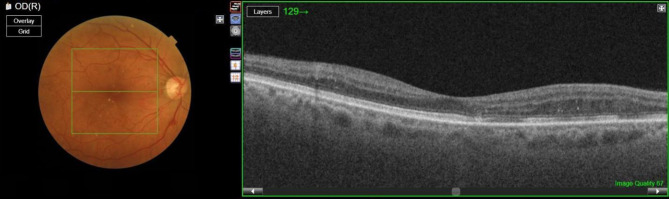
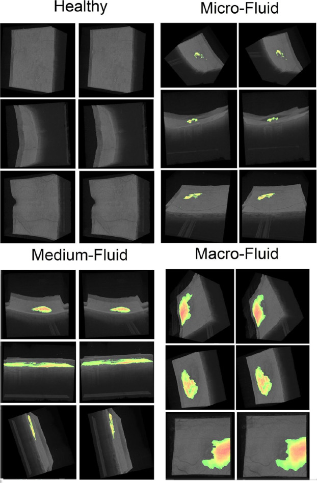
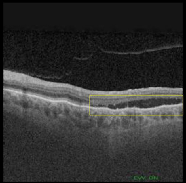
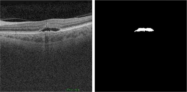
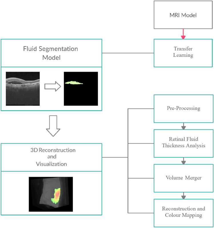
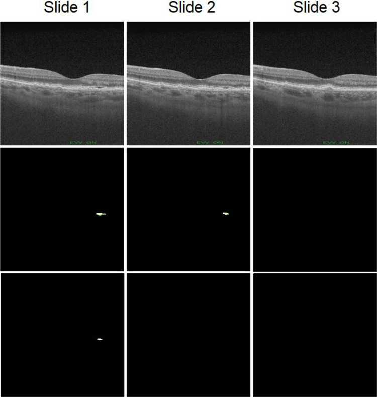
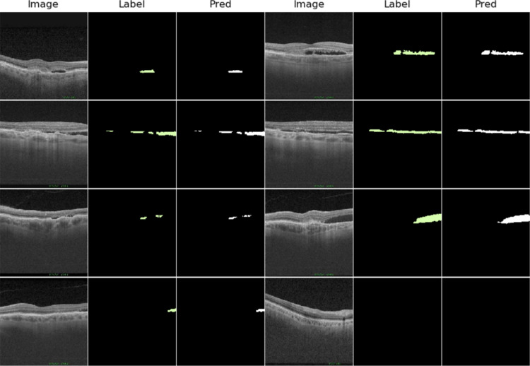
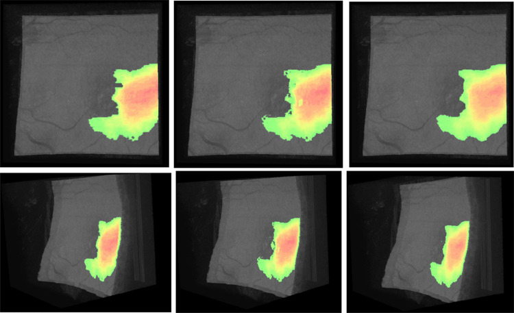
Age-related macular degeneration is the leading cause of vision loss in developed countries, and wet-type AMD requires urgent treatment and rapid diagnosis because it causes rapid irreversible vision loss. Currently, AMD diagnosis is mainly carried out using images obtained by optical coherence tomography. This diagnostic process is performed by human clinicians, so human error may occur in some cases. Therefore, fully automatic methodologies are highly desirable adding a layer of robustness to the diagnosis. In this work, a novel computer-aided diagnosis and visualization methodology is proposed for the rapid identification and visualization of wet AMD. We adapted a convolutional neural network for segmentation of a similar domain of medical images to the problem of wet AMD segmentation, taking advantage of transfer learning, which allows us to work with and exploit a reduced number of samples. We generate a 3D intuitive visualization where the existence, position and severity of the fluid were represented in a clear and intuitive way to facilitate the analysis of the clinicians. The 3D visualization is robust and accurate, obtaining satisfactory 0.949 and 0.960 Dice coefficients in the different evaluated OCT cube configurations, allowing to quickly assess the presence and extension of the fluid associated to wet AMD.

摘要

年龄相关性黄斑变性是发达国家视力丧失的主要原因,湿性 AMD 需要紧急治疗和快速诊断,因为它会导致迅速不可逆转的视力丧失。目前,AMD 的诊断主要是通过光学相干断层扫描获得的图像进行的。这个诊断过程是由人类临床医生进行的,因此在某些情况下可能会出现人为错误。因此,非常需要全自动的方法来为诊断增加一层稳健性。在这项工作中,我们提出了一种新的计算机辅助诊断和可视化方法,用于快速识别和可视化湿性 AMD。我们采用了卷积神经网络来分割与湿性 AMD 分割问题类似的医学图像领域,利用迁移学习,这使我们可以使用和利用较少的样本。我们生成了一个直观的 3D 可视化,以清晰直观的方式表示液体的存在、位置和严重程度,以方便临床医生进行分析。3D 可视化是稳健和准确的,在不同评估的 OCT 立方体配置中获得了令人满意的 0.949 和 0.960 的 Dice 系数,能够快速评估与湿性 AMD 相关的液体的存在和扩展。

https://cdn.ncbi.nlm.nih.gov/pmc/blobs/779d/9582110/b8b4f5608812/10278_2022_643_Fig11_HTML.jpg
https://cdn.ncbi.nlm.nih.gov/pmc/blobs/779d/9582110/368c01b59dfd/10278_2022_643_Fig1_HTML.jpg
https://cdn.ncbi.nlm.nih.gov/pmc/blobs/779d/9582110/e32164b6e619/10278_2022_643_Fig2_HTML.jpg
https://cdn.ncbi.nlm.nih.gov/pmc/blobs/779d/9582110/9b6155426ff9/10278_2022_643_Fig3_HTML.jpg
https://cdn.ncbi.nlm.nih.gov/pmc/blobs/779d/9582110/32efc1e02d01/10278_2022_643_Fig4_HTML.jpg
https://cdn.ncbi.nlm.nih.gov/pmc/blobs/779d/9582110/252423df260d/10278_2022_643_Fig5_HTML.jpg
https://cdn.ncbi.nlm.nih.gov/pmc/blobs/779d/9582110/6136139589b3/10278_2022_643_Fig6_HTML.jpg
https://cdn.ncbi.nlm.nih.gov/pmc/blobs/779d/9582110/f92357670119/10278_2022_643_Fig7_HTML.jpg
https://cdn.ncbi.nlm.nih.gov/pmc/blobs/779d/9582110/064375701adf/10278_2022_643_Fig8_HTML.jpg
https://cdn.ncbi.nlm.nih.gov/pmc/blobs/779d/9582110/38f4a6ce4531/10278_2022_643_Fig9_HTML.jpg
https://cdn.ncbi.nlm.nih.gov/pmc/blobs/779d/9582110/65d8605d9f35/10278_2022_643_Fig10_HTML.jpg
https://cdn.ncbi.nlm.nih.gov/pmc/blobs/779d/9582110/b8b4f5608812/10278_2022_643_Fig11_HTML.jpg

相似文献

1
Fully-Automatic 3D Intuitive Visualization of Age-Related Macular Degeneration Fluid Accumulations in OCT Cubes.OCT 立方体中与年龄相关的黄斑变性液体积聚的全自动 3D 直观可视化。
J Digit Imaging. 2022 Oct;35(5):1271-1282. doi: 10.1007/s10278-022-00643-6. Epub 2022 May 5.
2
Weakly supervised lesion localization for age-related macular degeneration detection using optical coherence tomography images.基于光学相干断层扫描图像的年龄相关性黄斑变性检测的弱监督病变定位。
PLoS One. 2019 Apr 5;14(4):e0215076. doi: 10.1371/journal.pone.0215076. eCollection 2019.
3
Automatic detection of retinal regions using fully convolutional networks for diagnosis of abnormal maculae in optical coherence tomography images.利用全卷积网络自动检测视网膜区域,用于光学相干断层扫描图像中黄斑异常的诊断。
J Biomed Opt. 2019 May;24(5):1-9. doi: 10.1117/1.JBO.24.5.056003.
4
U-Net-Based Segmentation of Current Imaging Biomarkers in OCT-Scans of Patients with Age Related Macular Degeneration.基于 U-Net 的年龄相关性黄斑变性患者 OCT 扫描中当前成像生物标志物的分割。
Stud Health Technol Inform. 2023 May 18;302:947-951. doi: 10.3233/SHTI230315.
5
Fully-automated atrophy segmentation in dry age-related macular degeneration in optical coherence tomography.光学相干断层扫描在干性年龄相关性黄斑变性中的全自动萎缩分割。
Sci Rep. 2021 Nov 8;11(1):21893. doi: 10.1038/s41598-021-01227-0.
6
Multi-scale convolutional neural network for automated AMD classification using retinal OCT images.用于使用视网膜光学相干断层扫描(OCT)图像进行年龄相关性黄斑变性(AMD)自动分类的多尺度卷积神经网络。
Comput Biol Med. 2022 May;144:105368. doi: 10.1016/j.compbiomed.2022.105368. Epub 2022 Mar 2.
7
Automatic segmentation of hyperreflective foci in OCT images.自动分割 OCT 图像中的高反射焦点。
Comput Methods Programs Biomed. 2019 Sep;178:91-103. doi: 10.1016/j.cmpb.2019.06.019. Epub 2019 Jun 17.
8
Deep learning to detect macular atrophy in wet age-related macular degeneration using optical coherence tomography.使用光学相干断层扫描的深度学习检测湿性年龄相关性黄斑变性的黄斑萎缩。
Sci Rep. 2023 May 22;13(1):8296. doi: 10.1038/s41598-023-35414-y.
9
The possibility of the combination of OCT and fundus images for improving the diagnostic accuracy of deep learning for age-related macular degeneration: a preliminary experiment.OCT 与眼底图像相结合提高深度学习诊断年龄相关性黄斑变性准确性的可能性:一项初步实验。
Med Biol Eng Comput. 2019 Mar;57(3):677-687. doi: 10.1007/s11517-018-1915-z. Epub 2018 Oct 22.
10
Understanding and interpreting CNN's decision in optical coherence tomography-based AMD detection.理解并解读卷积神经网络在基于光学相干断层扫描的 AMD 检测中的决策。
Eur J Ophthalmol. 2024 May;34(3):803-815. doi: 10.1177/11206721231199126. Epub 2023 Sep 6.

引用本文的文献

1
Artificial intelligence for diagnosing exudative age-related macular degeneration.人工智能在渗出性年龄相关性黄斑变性诊断中的应用。
Cochrane Database Syst Rev. 2024 Oct 17;10(10):CD015522. doi: 10.1002/14651858.CD015522.pub2.
2
Artificial intelligence in age-related macular degeneration: state of the art and recent updates.人工智能在年龄相关性黄斑变性中的应用:现状与最新进展。
BMC Ophthalmol. 2024 Mar 15;24(1):121. doi: 10.1186/s12886-024-03381-1.

本文引用的文献

1
3D Shape Modeling and Analysis of Retinal Microvasculature in OCT-Angiography Images.OCT 血管造影图像中视网膜微血管的 3D 形状建模与分析。
IEEE Trans Med Imaging. 2020 May;39(5):1335-1346. doi: 10.1109/TMI.2019.2948867. Epub 2019 Oct 22.
2
Association of genomic subtypes of lower-grade gliomas with shape features automatically extracted by a deep learning algorithm.低级别胶质瘤基因组亚型与深度学习算法自动提取的形态特征的关联。
Comput Biol Med. 2019 Jun;109:218-225. doi: 10.1016/j.compbiomed.2019.05.002. Epub 2019 May 3.
3
Deep-learning based multiclass retinal fluid segmentation and detection in optical coherence tomography images using a fully convolutional neural network.
基于深度学习的全卷积神经网络在光学相干断层扫描图像中对多类视网膜液的分割和检测。
Med Image Anal. 2019 May;54:100-110. doi: 10.1016/j.media.2019.02.011. Epub 2019 Feb 22.
4
OCT Fluid Segmentation using Graph Shortest Path and Convolutional Neural Network.基于图最短路径和卷积神经网络的光学相干断层扫描(OCT)流体分割
Annu Int Conf IEEE Eng Med Biol Soc. 2018 Jul;2018:3426-3429. doi: 10.1109/EMBC.2018.8512998.
5
Deep learning approach for the detection and quantification of intraretinal cystoid fluid in multivendor optical coherence tomography.用于在多厂商光学相干断层扫描中检测和定量视网膜内囊样液的深度学习方法。
Biomed Opt Express. 2018 Mar 7;9(4):1545-1569. doi: 10.1364/BOE.9.001545. eCollection 2018 Apr 1.
6
Deep-learning based, automated segmentation of macular edema in optical coherence tomography.基于深度学习的光学相干断层扫描中黄斑水肿的自动分割
Biomed Opt Express. 2017 Jun 23;8(7):3440-3448. doi: 10.1364/BOE.8.003440. eCollection 2017 Jul 1.
7
Volume-Rendered Optical Coherence Tomography of Diabetic Retinopathy Pilot Study.糖尿病视网膜病变的容积渲染光学相干断层扫描试点研究
Am J Ophthalmol. 2015 Dec;160(6):1200-10. doi: 10.1016/j.ajo.2015.09.010. Epub 2015 Sep 15.
8
Metrics for evaluating 3D medical image segmentation: analysis, selection, and tool.用于评估3D医学图像分割的指标:分析、选择与工具
BMC Med Imaging. 2015 Aug 12;15:29. doi: 10.1186/s12880-015-0068-x.
9
Global prevalence of age-related macular degeneration and disease burden projection for 2020 and 2040: a systematic review and meta-analysis.全球与年龄相关的黄斑变性患病率及 2020 与 2040 年疾病负担预测:系统回顾和荟萃分析。
Lancet Glob Health. 2014 Feb;2(2):e106-16. doi: 10.1016/S2214-109X(13)70145-1. Epub 2014 Jan 3.
10
Prevalence and causes of vision loss in high-income countries and in Eastern and Central Europe: 1990-2010.高收入国家以及东欧和中欧地区视力丧失的患病率及原因:1990 - 2010年
Br J Ophthalmol. 2014 May;98(5):629-38. doi: 10.1136/bjophthalmol-2013-304033. Epub 2014 Mar 24.