Tangeman Jared A, Pérez-Estrada J Raúl, Van Zeeland Emily, Liu Lin, Danciutiu Alexandra, Grajales-Esquivel Erika, Smucker Byran, Liang Chun, Del Rio-Tsonis Katia
Department of Biology and Center for Visual Sciences, Miami University, Oxford, OH, United States.
Department of Statistics, Miami University, Oxford, OH, United States.
Front Cell Dev Biol. 2022 Apr 19;10:875155. doi: 10.3389/fcell.2022.875155. eCollection 2022.
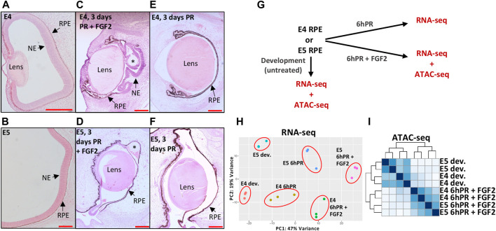
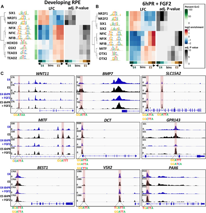
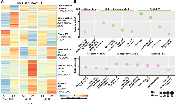
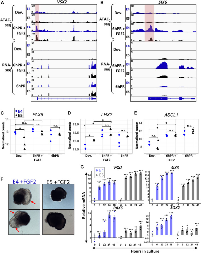
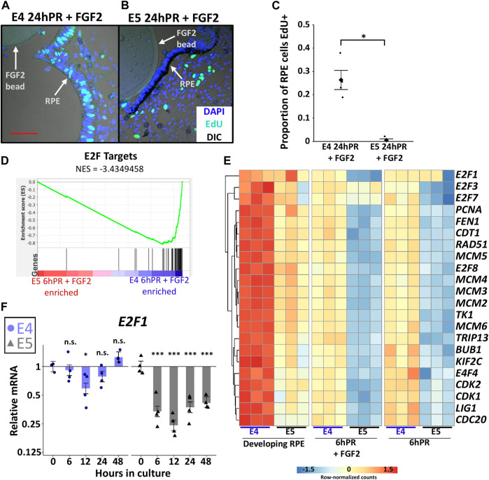
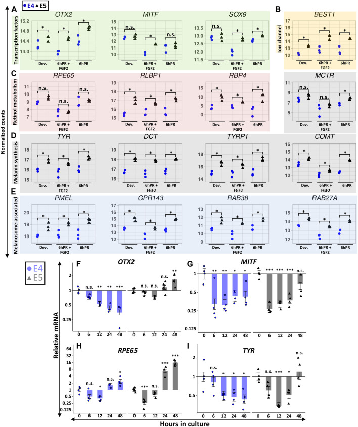
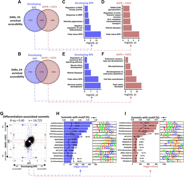
The retinal pigment epithelium (RPE) exhibits a diverse range of plasticity across vertebrates and is a potential source of cells for the regeneration of retinal neurons. Embryonic amniotes possess a transitory ability to regenerate neural retina through the reprogramming of RPE cells in an FGF-dependent manner. Chicken RPE can regenerate neural retina at embryonic day 4 (E4), but RPE neural competence is lost by embryonic day 5 (E5). To identify mechanisms that underlie loss of regenerative competence, we performed RNA and ATAC sequencing using E4 and E5 chicken RPE, as well as at both stages following retinectomy and FGF2 treatment. We find that genes associated with neural retina fate remain FGF2-inducible in the non-regenerative E5 RPE. Coinciding with fate restriction, RPE cells stably exit the cell cycle and dampen the expression of cell cycle progression genes normally expressed during regeneration, including . E5 RPE exhibits progressive activation of gene pathways associated with mature function independently of retinectomy or FGF2 treatment, including retinal metabolism, pigmentation synthesis, and ion transport. Moreover, the E5 RPE fails to efficiently repress expression in response to FGF2. Predicted OTX2 binding motifs undergo robust accessibility increases in E5 RPE, many of which coincide with putative regulatory elements for genes known to facilitate RPE differentiation and maturation. Together, these results uncover widespread alterations in gene regulation that culminate in the loss of RPE neural competence and implicate OTX2 as a key determinant in solidifying the RPE fate. These results yield valuable insight to the basis of RPE lineage restriction during early development and will be of importance in understanding the varying capacities for RPE-derived retinal regeneration observed among vertebrates.
视网膜色素上皮(RPE)在脊椎动物中表现出多种可塑性,是视网膜神经元再生的潜在细胞来源。胚胎羊膜动物具有通过FGF依赖的方式对RPE细胞进行重编程来再生神经视网膜的短暂能力。鸡的RPE在胚胎第4天(E4)可以再生神经视网膜,但在胚胎第5天(E5)时RPE的神经能力丧失。为了确定导致再生能力丧失的机制,我们使用E4和E5鸡的RPE,以及视网膜切除和FGF2处理后的两个阶段进行了RNA和ATAC测序。我们发现,与神经视网膜命运相关的基因在不具有再生能力的E5 RPE中仍可被FGF2诱导。与命运限制同时发生的是,RPE细胞稳定地退出细胞周期,并抑制再生过程中正常表达的细胞周期进展基因的表达,包括 。E5 RPE表现出与成熟功能相关的基因途径的渐进激活,这与视网膜切除或FGF2处理无关,包括视网膜代谢、色素合成和离子转运。此外,E5 RPE不能有效地响应FGF2抑制 表达。预测的OTX2结合基序在E5 RPE中可及性显著增加,其中许多与已知促进RPE分化和成熟的基因的假定调控元件一致。总之,这些结果揭示了基因调控中广泛的变化,这些变化最终导致RPE神经能力的丧失,并表明OTX2是巩固RPE命运的关键决定因素。这些结果为早期发育过程中RPE谱系限制的基础提供了有价值的见解,对于理解脊椎动物中观察到的RPE衍生的视网膜再生的不同能力具有重要意义。