• 文献检索
  • 文档翻译
  • 深度研究
  • 学术资讯
  • Suppr Zotero 插件Zotero 插件
  • 邀请有礼
  • 套餐&价格
  • 历史记录
应用&插件
Suppr Zotero 插件Zotero 插件浏览器插件Mac 客户端Windows 客户端微信小程序
定价
高级版会员购买积分包购买API积分包
服务
文献检索文档翻译深度研究API 文档MCP 服务
关于我们
关于 Suppr公司介绍联系我们用户协议隐私条款
关注我们

Suppr 超能文献

核心技术专利:CN118964589B侵权必究
粤ICP备2023148730 号-1Suppr @ 2026

文献检索

告别复杂PubMed语法,用中文像聊天一样搜索,搜遍4000万医学文献。AI智能推荐,让科研检索更轻松。

立即免费搜索

文件翻译

保留排版,准确专业,支持PDF/Word/PPT等文件格式,支持 12+语言互译。

免费翻译文档

深度研究

AI帮你快速写综述,25分钟生成高质量综述,智能提取关键信息,辅助科研写作。

立即免费体验

光学显微镜中基于深度学习的图像处理

Deep learning-based image processing in optical microscopy.

作者信息

Melanthota Sindhoora Kaniyala, Gopal Dharshini, Chakrabarti Shweta, Kashyap Anirudh Ameya, Radhakrishnan Raghu, Mazumder Nirmal

机构信息

Department of Biophysics, Manipal School of Life Sciences, Manipal Academy of Higher Education, Manipal, Karnataka 576104 India.

Department of Bioinformatics, Manipal School of Life Sciences, Manipal Academy of Higher Education, Manipal, Karnataka 576104 India.

出版信息

Biophys Rev. 2022 Apr 6;14(2):463-481. doi: 10.1007/s12551-022-00949-3. eCollection 2022 Apr.

DOI:10.1007/s12551-022-00949-3
PMID:35528030
原文链接:https://pmc.ncbi.nlm.nih.gov/articles/PMC9043085/
Abstract

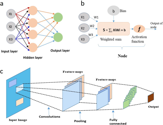
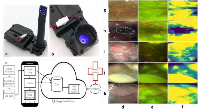
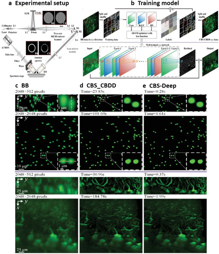
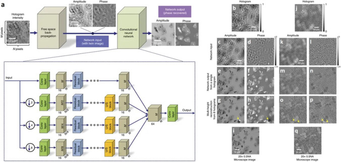
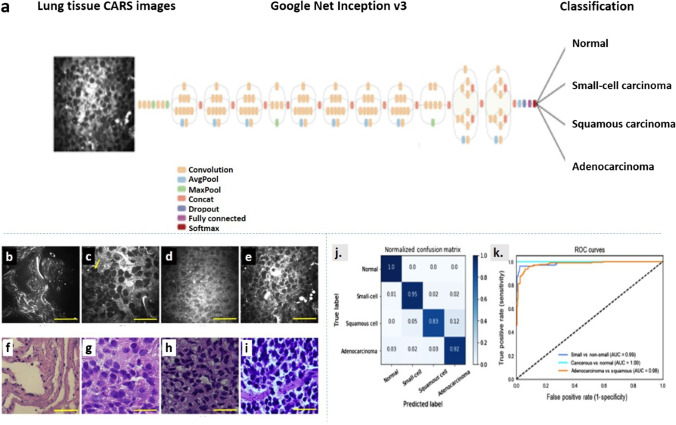
Optical microscopy has emerged as a key driver of fundamental research since it provides the ability to probe into imperceptible structures in the biomedical world. For the detailed investigation of samples, a high-resolution image with enhanced contrast and minimal damage is preferred. To achieve this, an automated image analysis method is preferable over manual analysis in terms of both speed of acquisition and reduced error accumulation. In this regard, deep learning (DL)-based image processing can be highly beneficial. The review summarises and critiques the use of DL in image processing for the data collected using various optical microscopic techniques. In tandem with optical microscopy, DL has already found applications in various problems related to image classification and segmentation. It has also performed well in enhancing image resolution in smartphone-based microscopy, which in turn enablse crucial medical assistance in remote places.

摘要

光学显微镜已成为基础研究的关键驱动力,因为它能够探测生物医学领域中难以察觉的结构。对于样品的详细研究,人们更倾向于具有增强对比度和最小损伤的高分辨率图像。为此,在采集速度和减少误差积累方面,自动图像分析方法优于手动分析。在这方面,基于深度学习(DL)的图像处理可能非常有益。这篇综述总结并评论了深度学习在图像处理中的应用,这些图像数据是使用各种光学显微镜技术收集的。与光学显微镜相结合,深度学习已经在与图像分类和分割相关的各种问题中得到应用。它在提高基于智能手机的显微镜的图像分辨率方面也表现出色,这反过来又能在偏远地区提供关键的医疗帮助。

https://cdn.ncbi.nlm.nih.gov/pmc/blobs/1929/9043085/9f71b1d60717/12551_2022_949_Fig8_HTML.jpg
https://cdn.ncbi.nlm.nih.gov/pmc/blobs/1929/9043085/29817139ca3a/12551_2022_949_Fig1_HTML.jpg
https://cdn.ncbi.nlm.nih.gov/pmc/blobs/1929/9043085/20ea757aa335/12551_2022_949_Fig2_HTML.jpg
https://cdn.ncbi.nlm.nih.gov/pmc/blobs/1929/9043085/ef965c9901ba/12551_2022_949_Fig3_HTML.jpg
https://cdn.ncbi.nlm.nih.gov/pmc/blobs/1929/9043085/0ea0f89e91e0/12551_2022_949_Fig4_HTML.jpg
https://cdn.ncbi.nlm.nih.gov/pmc/blobs/1929/9043085/684f5a82c6c3/12551_2022_949_Fig5_HTML.jpg
https://cdn.ncbi.nlm.nih.gov/pmc/blobs/1929/9043085/2d997b4c44bc/12551_2022_949_Fig6_HTML.jpg
https://cdn.ncbi.nlm.nih.gov/pmc/blobs/1929/9043085/90e07a07b83a/12551_2022_949_Fig7_HTML.jpg
https://cdn.ncbi.nlm.nih.gov/pmc/blobs/1929/9043085/9f71b1d60717/12551_2022_949_Fig8_HTML.jpg

相似文献

1
Deep learning-based image processing in optical microscopy.光学显微镜中基于深度学习的图像处理
Biophys Rev. 2022 Apr 6;14(2):463-481. doi: 10.1007/s12551-022-00949-3. eCollection 2022 Apr.
2
Portable, Automated and Deep-Learning-Enabled Microscopy for Smartphone-Tethered Optical Platform Towards Remote Homecare Diagnostics: A Review.用于智能手机连接光学平台的便携式、自动化和深度学习显微镜在远程家庭护理诊断中的应用:综述
Small Methods. 2023 Jan;7(1):e2200979. doi: 10.1002/smtd.202200979. Epub 2022 Nov 24.
3
Deep Learning-Based Single-Cell Optical Image Studies.基于深度学习的单细胞光学图像研究。
Cytometry A. 2020 Mar;97(3):226-240. doi: 10.1002/cyto.a.23973. Epub 2020 Jan 25.
4
Design of a 3D printed smartphone microscopic system with enhanced imaging ability for biomedical applications.用于生物医学应用的具有增强成像能力的 3D 打印智能手机显微镜系统的设计。
J Microsc. 2019 Oct;276(1):13-20. doi: 10.1111/jmi.12829. Epub 2019 Sep 22.
5
Automated Microscopy Image Segmentation and Analysis with Machine Learning.基于机器学习的自动化显微镜图像分割与分析。
Methods Mol Biol. 2022;2440:349-365. doi: 10.1007/978-1-0716-2051-9_20.
6
A survey on applications of deep learning in microscopy image analysis.深度学习在显微镜图像分析中的应用调查。
Comput Biol Med. 2021 Jul;134:104523. doi: 10.1016/j.compbiomed.2021.104523. Epub 2021 May 29.
7
Tile-based microscopic image processing for malaria screening using a deep learning approach.基于瓦片的显微镜图像深度学习处理在疟疾筛查中的应用。
BMC Med Imaging. 2023 Mar 22;23(1):39. doi: 10.1186/s12880-023-00993-9.
8
Deep learning provides high accuracy in automated chondrocyte viability assessment in articular cartilage using nonlinear optical microscopy.深度学习在使用非线性光学显微镜对关节软骨中的软骨细胞活力进行自动评估时具有很高的准确性。
Biomed Opt Express. 2021 Apr 16;12(5):2759-2772. doi: 10.1364/BOE.417478. eCollection 2021 May 1.
9
Unraveling the complexity of Optical Coherence Tomography image segmentation using machine and deep learning techniques: A review.利用机器学习和深度学习技术解析光学相干断层扫描图像分割的复杂性:综述。
Comput Med Imaging Graph. 2023 Sep;108:102269. doi: 10.1016/j.compmedimag.2023.102269. Epub 2023 Jul 14.
10
Generative and discriminative model-based approaches to microscopic image restoration and segmentation.基于生成模型和判别模型的微观图像恢复与分割方法。
Microscopy (Oxf). 2020 Apr 8;69(2):79-91. doi: 10.1093/jmicro/dfaa007.

引用本文的文献

1
Enhancing AI microscopy for foodborne bacterial classification using adversarial domain adaptation to address optical and biological variability.利用对抗域适应增强人工智能显微镜用于食源细菌分类,以解决光学和生物学变异性问题。
Front Artif Intell. 2025 Aug 12;8:1632344. doi: 10.3389/frai.2025.1632344. eCollection 2025.
2
Advancing Multicolor Super-Resolution Volume Imaging: Illuminating Complex Cellular Dynamics.推进多色超分辨率体积成像:揭示复杂的细胞动力学。
JACS Au. 2025 Jun 9;5(6):2388-2419. doi: 10.1021/jacsau.5c00314. eCollection 2025 Jun 23.
3
Cyber-physical security of biochips: A perspective.

本文引用的文献

1
Deep learning-enhanced light-field imaging with continuous validation.深度学习增强的具有连续验证的光场成像。
Nat Methods. 2021 May;18(5):557-563. doi: 10.1038/s41592-021-01136-0. Epub 2021 May 7.
2
Deep learning-enabled efficient image restoration for 3D microscopy of turbid biological specimens.深度学习助力混浊生物样本三维显微镜成像的高效图像恢复。
Opt Express. 2020 Sep 28;28(20):30234-30247. doi: 10.1364/OE.399542.
3
Deep learning-based cell identification and disease diagnosis using spatio-temporal cellular dynamics in compact digital holographic microscopy.
生物芯片的网络物理安全:一种视角。
Biomicrofluidics. 2025 May 29;19(3):031304. doi: 10.1063/5.0252554. eCollection 2025 May.
4
Microscope Upcycling: Transforming legacy microscopes into automated cloud-integrated imaging systems.显微镜升级改造:将传统显微镜转变为自动化云集成成像系统。
HardwareX. 2025 Mar 19;22:e00637. doi: 10.1016/j.ohx.2025.e00637. eCollection 2025 Jun.
5
A novel method (RIM-Deep) for enhancing imaging depth and resolution stability of deep cleared tissue in inverted confocal microscopy.一种用于提高倒置共聚焦显微镜中深度清除组织成像深度和分辨率稳定性的新方法(RIM-Deep)。
Elife. 2025 Apr 7;13:RP101143. doi: 10.7554/eLife.101143.
6
Applications of Artificial Intelligence, Deep Learning, and Machine Learning to Support the Analysis of Microscopic Images of Cells and Tissues.人工智能、深度学习和机器学习在支持细胞与组织微观图像分析中的应用。
J Imaging. 2025 Feb 15;11(2):59. doi: 10.3390/jimaging11020059.
7
Quantifying Monomer-Dimer Distribution of Nanoparticles from Uncorrelated Optical Images Using Deep Learning.使用深度学习从非相关光学图像中量化纳米颗粒的单体-二聚体分布
ACS Omega. 2025 Jan 3;10(1):862-870. doi: 10.1021/acsomega.4c07914. eCollection 2025 Jan 14.
8
Effect of shear rate on early adhesion dynamics monitored by deep learning.剪切速率对通过深度学习监测的早期黏附动力学的影响。
Biofilm. 2024 Nov 16;8:100240. doi: 10.1016/j.bioflm.2024.100240. eCollection 2024 Dec.
9
Image processing tools for petabyte-scale light sheet microscopy data.用于拍字节级光片显微镜数据的图像处理工具。
Nat Methods. 2024 Dec;21(12):2342-2352. doi: 10.1038/s41592-024-02475-4. Epub 2024 Oct 17.
10
Ex vivo nanoscale abluminal mapping of putative cargo receptors at the blood-brain barrier of expanded brain capillaries.体外扩张脑毛细血管血脑屏障中潜在货物受体的纳米级向腔侧绘图。
Fluids Barriers CNS. 2024 Oct 14;21(1):80. doi: 10.1186/s12987-024-00585-x.
在紧凑型数字全息显微镜中,基于深度学习利用时空细胞动力学进行细胞识别和疾病诊断。
Biomed Opt Express. 2020 Jul 21;11(8):4491-4508. doi: 10.1364/BOE.399020. eCollection 2020 Aug 1.
4
Noise-free quantitative phase imaging in Gabor holography with conditional generative adversarial network.基于条件生成对抗网络的加博尔全息术中的无噪声定量相位成像。
Opt Express. 2020 Aug 31;28(18):26284-26301. doi: 10.1364/OE.398528.
5
High spatially sensitive quantitative phase imaging assisted with deep neural network for classification of human spermatozoa under stressed condition.高空间灵敏度定量相位成像辅助深度神经网络对应激状态下人精子的分类。
Sci Rep. 2020 Aug 4;10(1):13118. doi: 10.1038/s41598-020-69857-4.
6
Deep learning-enhanced fluorescence microscopy via degeneration decoupling.通过退化解耦实现深度学习增强荧光显微镜技术
Opt Express. 2020 May 11;28(10):14859-14873. doi: 10.1364/OE.390121.
7
Resolving challenges in deep learning-based analyses of histopathological images using explanation methods.运用解释方法解决基于深度学习的组织病理学图像分析中的挑战。
Sci Rep. 2020 Apr 14;10(1):6423. doi: 10.1038/s41598-020-62724-2.
8
Deep learning a boon for biophotonics?深度学习对生物光子学是福音吗?
J Biophotonics. 2020 Jun;13(6):e201960186. doi: 10.1002/jbio.201960186. Epub 2020 Mar 30.
9
Applications of Deep Learning for Dense Scenes Analysis in Agriculture: A Review.深度学习在农业密集场景分析中的应用综述。
Sensors (Basel). 2020 Mar 10;20(5):1520. doi: 10.3390/s20051520.
10
Three-dimensional virtual refocusing of fluorescence microscopy images using deep learning.基于深度学习的荧光显微镜图像三维虚拟聚焦。
Nat Methods. 2019 Dec;16(12):1323-1331. doi: 10.1038/s41592-019-0622-5. Epub 2019 Nov 4.