• 文献检索
  • 文档翻译
  • 深度研究
  • 学术资讯
  • Suppr Zotero 插件Zotero 插件
  • 邀请有礼
  • 套餐&价格
  • 历史记录
应用&插件
Suppr Zotero 插件Zotero 插件浏览器插件Mac 客户端Windows 客户端微信小程序
定价
高级版会员购买积分包购买API积分包
服务
文献检索文档翻译深度研究API 文档MCP 服务
关于我们
关于 Suppr公司介绍联系我们用户协议隐私条款
关注我们

Suppr 超能文献

核心技术专利:CN118964589B侵权必究
粤ICP备2023148730 号-1Suppr @ 2026

文献检索

告别复杂PubMed语法,用中文像聊天一样搜索,搜遍4000万医学文献。AI智能推荐,让科研检索更轻松。

立即免费搜索

文件翻译

保留排版,准确专业,支持PDF/Word/PPT等文件格式,支持 12+语言互译。

免费翻译文档

深度研究

AI帮你快速写综述,25分钟生成高质量综述,智能提取关键信息,辅助科研写作。

立即免费体验

COVID-19 及其他病理性冠状病毒的胸部影像学表现综述。

Review of Thoracic Imaging Manifestations of COVID-19 and Other Pathologic Coronaviruses.

机构信息

Department of Diagnostic, Molecular and Interventional Radiology, Icahn School of Medicine at Mount Sinai, Mount Sinai Hospital, New York, NY 10029, USA.

Department of Diagnostic, Molecular and Interventional Radiology, Icahn School of Medicine at Mount Sinai, Mount Sinai Hospital, New York, NY 10029, USA.

出版信息

Radiol Clin North Am. 2022 May;60(3):359-369. doi: 10.1016/j.rcl.2022.01.004. Epub 2022 Jan 11.

DOI:10.1016/j.rcl.2022.01.004
PMID:35534124
原文链接:https://pmc.ncbi.nlm.nih.gov/articles/PMC8747969/
Abstract

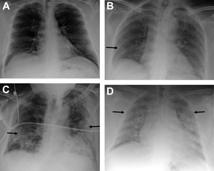
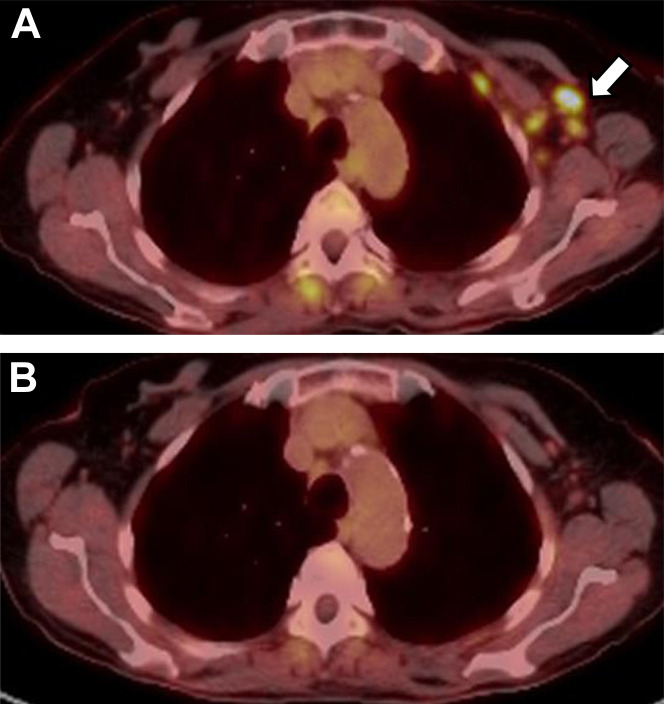
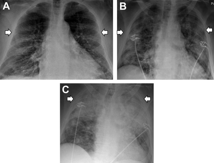
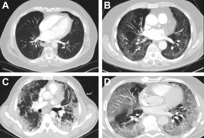
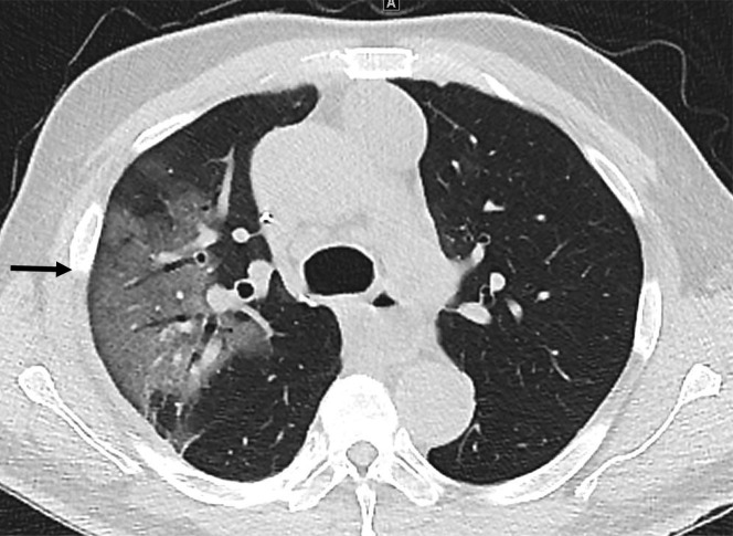
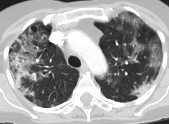
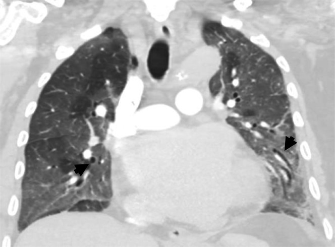
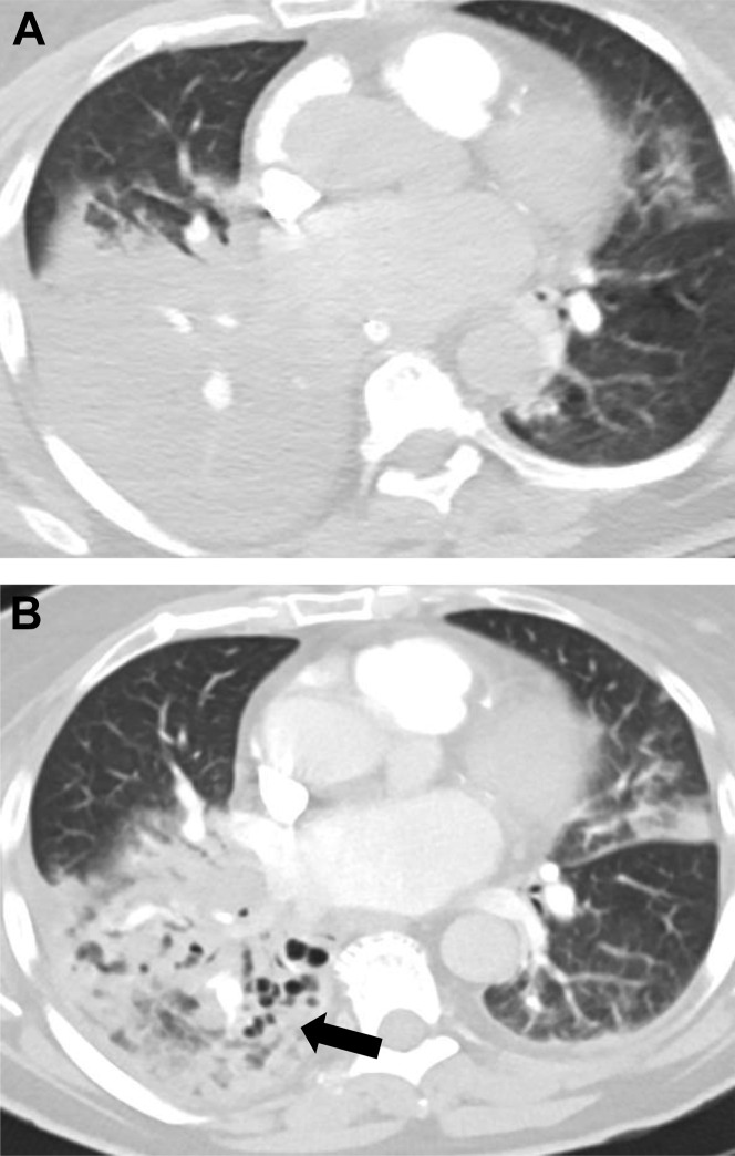
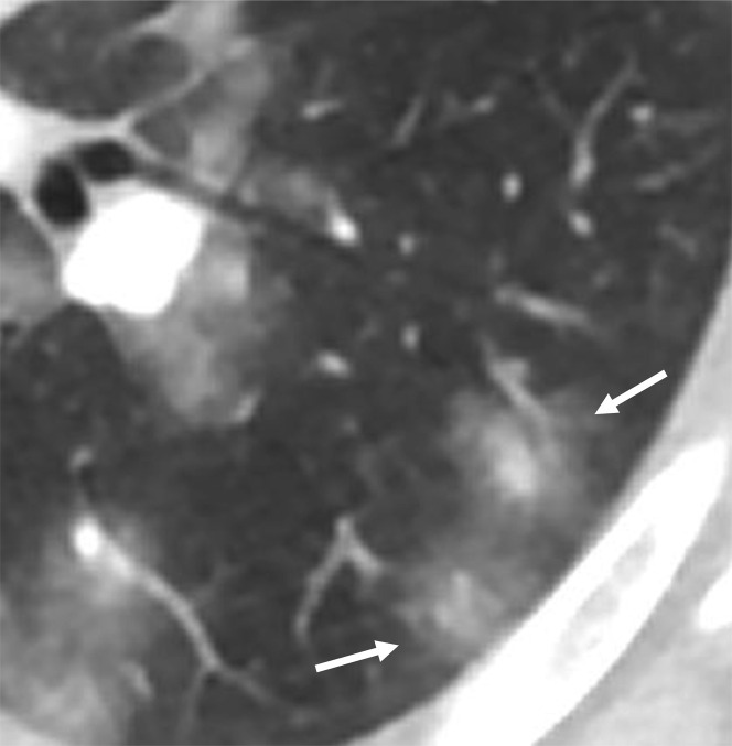
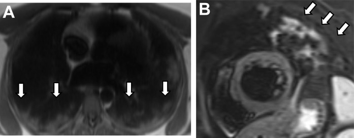
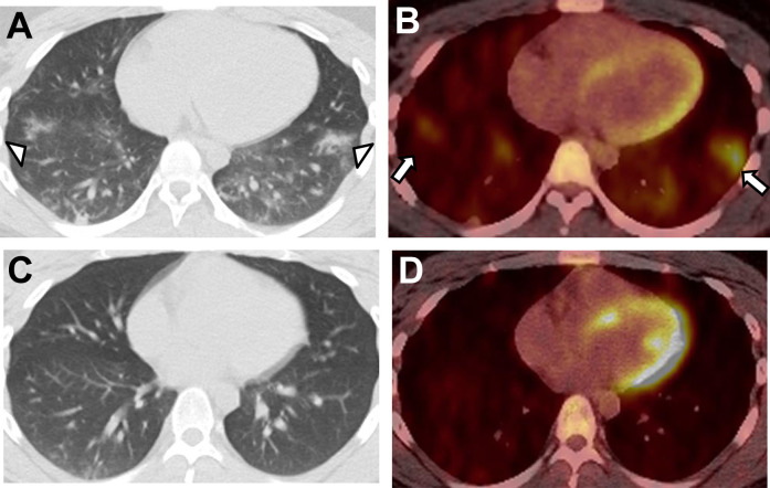
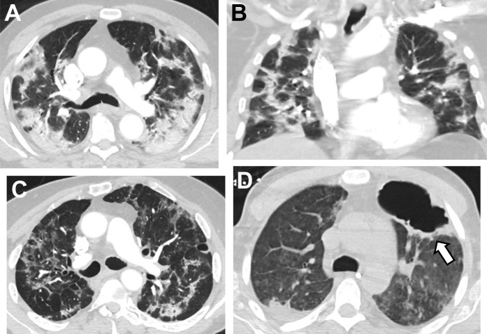
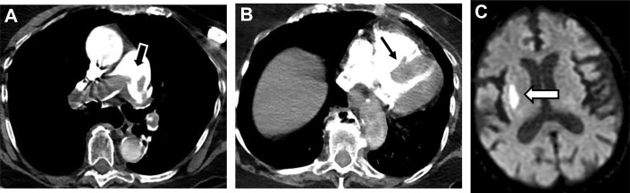
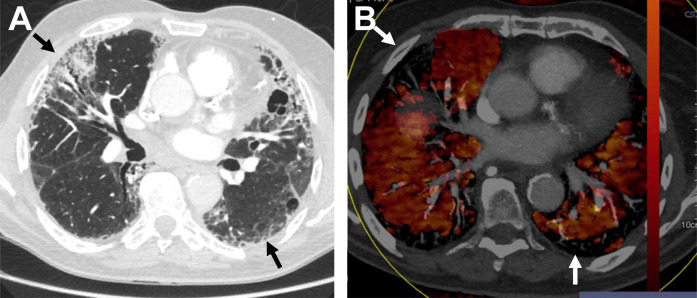
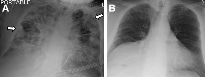
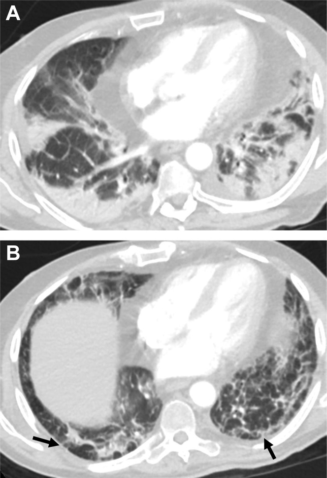
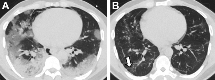
Severe acute respiratory syndrome coronavirus 2 (SARS-CoV-2) is an easily transmissible coronavirus that emerged in late 2019 and has caused a global pandemic characterized by acute respiratory disease named coronavirus disease 2019 (COVID-19). Diagnostic imaging can be helpful as a complementary tool in supporting the diagnosis of COVID-19 and identifying alternative pathology. This article presents an overview of acute and postacute imaging findings in COVID-19.

摘要

严重急性呼吸综合征冠状病毒 2(SARS-CoV-2)是一种易传播的冠状病毒,于 2019 年末出现,并导致以急性呼吸道疾病命名的 2019 年冠状病毒病(COVID-19)为特征的全球大流行。诊断性影像学检查可以作为一种辅助手段,有助于支持 COVID-19 的诊断并识别其他病变。本文概述了 COVID-19 的急性和亚急性影像学表现。

https://cdn.ncbi.nlm.nih.gov/pmc/blobs/d87b/8747969/1e1e2760baa7/gr18_lrg.jpg
https://cdn.ncbi.nlm.nih.gov/pmc/blobs/d87b/8747969/04be76a9e0f1/gr1_lrg.jpg
https://cdn.ncbi.nlm.nih.gov/pmc/blobs/d87b/8747969/9ce3b0902428/gr2_lrg.jpg
https://cdn.ncbi.nlm.nih.gov/pmc/blobs/d87b/8747969/ab45dd60bfa8/gr3_lrg.jpg
https://cdn.ncbi.nlm.nih.gov/pmc/blobs/d87b/8747969/274c99278940/gr4_lrg.jpg
https://cdn.ncbi.nlm.nih.gov/pmc/blobs/d87b/8747969/dcf7c4dc9a8d/gr5_lrg.jpg
https://cdn.ncbi.nlm.nih.gov/pmc/blobs/d87b/8747969/a29a36a973b8/gr6_lrg.jpg
https://cdn.ncbi.nlm.nih.gov/pmc/blobs/d87b/8747969/e99a5786304d/gr7_lrg.jpg
https://cdn.ncbi.nlm.nih.gov/pmc/blobs/d87b/8747969/1a20d39020a4/gr8_lrg.jpg
https://cdn.ncbi.nlm.nih.gov/pmc/blobs/d87b/8747969/2534931eb058/gr9_lrg.jpg
https://cdn.ncbi.nlm.nih.gov/pmc/blobs/d87b/8747969/01ee616765b8/gr10_lrg.jpg
https://cdn.ncbi.nlm.nih.gov/pmc/blobs/d87b/8747969/a7c0f27a2568/gr11_lrg.jpg
https://cdn.ncbi.nlm.nih.gov/pmc/blobs/d87b/8747969/a1db575da0a2/gr12_lrg.jpg
https://cdn.ncbi.nlm.nih.gov/pmc/blobs/d87b/8747969/49e02124baa6/gr13_lrg.jpg
https://cdn.ncbi.nlm.nih.gov/pmc/blobs/d87b/8747969/7995f6c74c03/gr14_lrg.jpg
https://cdn.ncbi.nlm.nih.gov/pmc/blobs/d87b/8747969/6f5b9375c9c9/gr15_lrg.jpg
https://cdn.ncbi.nlm.nih.gov/pmc/blobs/d87b/8747969/3d8eace34d60/gr16_lrg.jpg
https://cdn.ncbi.nlm.nih.gov/pmc/blobs/d87b/8747969/0d51136ff6d1/gr17_lrg.jpg
https://cdn.ncbi.nlm.nih.gov/pmc/blobs/d87b/8747969/1e1e2760baa7/gr18_lrg.jpg

相似文献

1
Review of Thoracic Imaging Manifestations of COVID-19 and Other Pathologic Coronaviruses.COVID-19 及其他病理性冠状病毒的胸部影像学表现综述。
Radiol Clin North Am. 2022 May;60(3):359-369. doi: 10.1016/j.rcl.2022.01.004. Epub 2022 Jan 11.
2
Human and novel coronavirus infections in children: a review.儿童人感染和新型冠状病毒感染:综述。
Paediatr Int Child Health. 2021 Feb;41(1):36-55. doi: 10.1080/20469047.2020.1781356. Epub 2020 Jun 25.
3
Coronaviruses - a new old menace.冠状病毒——新的旧威胁。
Postepy Biochem. 2021 Jan 5;66(4):303-308. doi: 10.18388/pb.2020_357. Print 2020 Dec 31.
4
[Diagnosis, treatment, control and prevention of SARS-CoV-2 and coronavirus disease 2019: back to the future].[2019冠状病毒病及严重急性呼吸综合征冠状病毒2的诊断、治疗、控制与预防:回归未来]
Sheng Wu Gong Cheng Xue Bao. 2020 Apr 25;36(4):571-592. doi: 10.13345/j.cjb.200115.
5
The unique features of SARS-CoV-2 transmission: Comparison with SARS-CoV, MERS-CoV and 2009 H1N1 pandemic influenza virus.SARS-CoV-2 传播的独特特征:与 SARS-CoV、MERS-CoV 和 2009 年 H1N1 大流行性流感病毒的比较。
Rev Med Virol. 2021 Mar;31(2):e2171. doi: 10.1002/rmv.2171. Epub 2020 Sep 18.
6
A comparative overview of COVID-19, MERS and SARS: Review article.新型冠状病毒病、中东呼吸综合征和严重急性呼吸综合征的比较概述:综述文章。
Int J Surg. 2020 Sep;81:1-8. doi: 10.1016/j.ijsu.2020.07.032. Epub 2020 Jul 26.
7
Biological, clinical and epidemiological features of COVID-19, SARS and MERS and AutoDock simulation of ACE2.新型冠状病毒肺炎、严重急性呼吸综合征和中东呼吸综合征的生物学、临床和流行病学特征,以及 ACE2 的 AutoDock 模拟
Infect Dis Poverty. 2020 Jul 20;9(1):99. doi: 10.1186/s40249-020-00691-6.
8
COVID-19: A Multidisciplinary Review.新型冠状病毒肺炎(COVID-19):多学科综述。
Front Public Health. 2020 Jul 29;8:383. doi: 10.3389/fpubh.2020.00383. eCollection 2020.
9
Highly pathogenic coronaviruses and the kidney.高致病性冠状病毒与肾脏。
Biomed Pharmacother. 2022 Dec;156:113807. doi: 10.1016/j.biopha.2022.113807. Epub 2022 Oct 11.
10
Recent Patents and Advances on Nanotechnologies against Coronavirus.近期冠状病毒纳米技术专利及进展
Recent Pat Nanotechnol. 2021;15(4):322-330. doi: 10.2174/1872210514666201110125536.

本文引用的文献

1
CT of Post-Acute Lung Complications of COVID-19.COVID-19 后肺部并发症的 CT 表现。
Radiology. 2021 Nov;301(2):E383-E395. doi: 10.1148/radiol.2021211396. Epub 2021 Aug 10.
2
Post-Acute Sequelae of COVID-19 Pneumonia: Six-month Chest CT Follow-up.新型冠状病毒肺炎的急性后遗症:胸部CT六个月随访
Radiology. 2021 Nov;301(2):E396-E405. doi: 10.1148/radiol.2021210834. Epub 2021 Jul 27.
3
Dual-Energy CT Pulmonary Angiography (DECTPA) Quantifies Vasculopathy in Severe COVID-19 Pneumonia.双能量CT肺血管造影(DECTPA)对重症新型冠状病毒肺炎血管病变进行定量分析。
Radiol Cardiothorac Imaging. 2020 Oct 29;2(5):e200428. doi: 10.1148/ryct.2020200428. eCollection 2020 Oct.
4
COVID-19: A Multimodality Review of Radiologic Techniques, Clinical Utility, and Imaging Features.新型冠状病毒肺炎:放射学技术、临床应用及影像特征的多模态综述
Radiol Cardiothorac Imaging. 2020 Jun 1;2(3):e200210. doi: 10.1148/ryct.2020200210. eCollection 2020 Jun.
5
Radiological Society of North America Expert Consensus Document on Reporting Chest CT Findings Related to COVID-19: Endorsed by the Society of Thoracic Radiology, the American College of Radiology, and RSNA.北美放射学会关于报告与COVID-19相关的胸部CT检查结果的专家共识文件:得到了胸腔放射学会、美国放射学会和北美放射学会的认可。
Radiol Cardiothorac Imaging. 2020 Mar 25;2(2):e200152. doi: 10.1148/ryct.2020200152. eCollection 2020 Apr.
6
Coronavirus Disease 2019: Initial Detection on Chest CT in a Retrospective Multicenter Study of 103 Chinese Patients.2019冠状病毒病:103例中国患者回顾性多中心研究中胸部CT的初步检测
Radiol Cardiothorac Imaging. 2020 Apr 6;2(2):e200092. doi: 10.1148/ryct.2020200092. eCollection 2020 Apr.
7
Chest CT Severity Score: An Imaging Tool for Assessing Severe COVID-19.胸部CT严重程度评分:一种评估重症COVID-19的影像学工具。
Radiol Cardiothorac Imaging. 2020 Mar 30;2(2):e200047. doi: 10.1148/ryct.2020200047. eCollection 2020 Apr.
8
Multidisciplinary Recommendations Regarding Post-Vaccine Adenopathy and Radiologic Imaging: Scientific Expert Panel.疫苗接种后淋巴结病和影像学检查的多学科建议:科学专家小组。
Radiology. 2021 Aug;300(2):E323-E327. doi: 10.1148/radiol.2021210436. Epub 2021 Feb 24.
9
Six-month Follow-up Chest CT Findings after Severe COVID-19 Pneumonia.重症 COVID-19 肺炎患者 6 个月随访的胸部 CT 结果。
Radiology. 2021 Apr;299(1):E177-E186. doi: 10.1148/radiol.2021203153. Epub 2021 Jan 26.
10
Unilateral axillary Adenopathy in the setting of COVID-19 vaccine.COVID-19 疫苗接种后出现单侧腋窝淋巴结病。
Clin Imaging. 2021 Jul;75:12-15. doi: 10.1016/j.clinimag.2021.01.016. Epub 2021 Jan 19.