• 文献检索
  • 文档翻译
  • 深度研究
  • 学术资讯
  • Suppr Zotero 插件Zotero 插件
  • 邀请有礼
  • 套餐&价格
  • 历史记录
应用&插件
Suppr Zotero 插件Zotero 插件浏览器插件Mac 客户端Windows 客户端微信小程序
定价
高级版会员购买积分包购买API积分包
服务
文献检索文档翻译深度研究API 文档MCP 服务
关于我们
关于 Suppr公司介绍联系我们用户协议隐私条款
关注我们

Suppr 超能文献

核心技术专利:CN118964589B侵权必究
粤ICP备2023148730 号-1Suppr @ 2026

文献检索

告别复杂PubMed语法,用中文像聊天一样搜索,搜遍4000万医学文献。AI智能推荐,让科研检索更轻松。

立即免费搜索

文件翻译

保留排版,准确专业,支持PDF/Word/PPT等文件格式,支持 12+语言互译。

免费翻译文档

深度研究

AI帮你快速写综述,25分钟生成高质量综述,智能提取关键信息,辅助科研写作。

立即免费体验

新型冠状病毒肺炎:放射学技术、临床应用及影像特征的多模态综述

COVID-19: A Multimodality Review of Radiologic Techniques, Clinical Utility, and Imaging Features.

作者信息

Manna Sayan, Wruble Jill, Maron Samuel Z, Toussie Danielle, Voutsinas Nicholas, Finkelstein Mark, Cedillo Mario A, Diamond Jamie, Eber Corey, Jacobi Adam, Chung Michael, Bernheim Adam

机构信息

Department of Diagnostic, Molecular and Interventional Radiology, Icahn School of Medicine at Mount Sinai, Mount Sinai Hospital, 1 Gustave L. Levy Place, New York, NY 10029 (S.M., S.Z.M., D.T., N.V., M.F., M.A.C., C.E., A.J., M.C., A.B.); Complete Radiology Reading Services, Westbury, NY (J.W.); Department of Radiology and Biomedical Imaging, Yale School of Medicine, New Haven, Conn (J.W.); Department of Radiology, University of Connecticut, Farmington, Conn (J.W.); Department of Radiology and Biomedical Imaging, The Johns Hopkins Hospital, Baltimore, Md (J.W.); and Division of Cardiology, Beth Israel Deaconess Medical Center, Boston, Mass (J.D.).

出版信息

Radiol Cardiothorac Imaging. 2020 Jun 1;2(3):e200210. doi: 10.1148/ryct.2020200210. eCollection 2020 Jun.

DOI:10.1148/ryct.2020200210
PMID:33778588
原文链接:https://pmc.ncbi.nlm.nih.gov/articles/PMC7325394/
Abstract

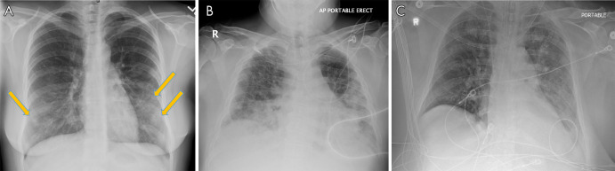
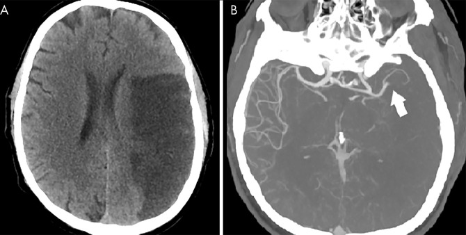
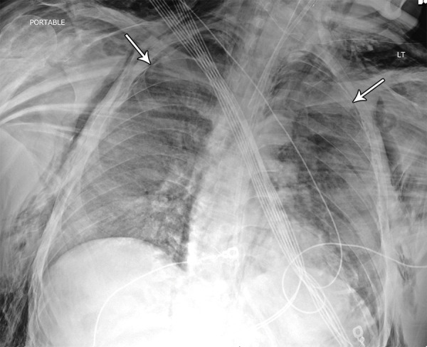
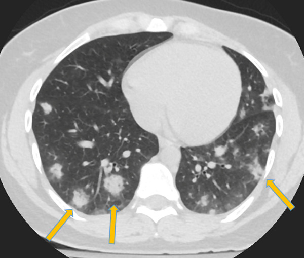
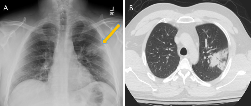
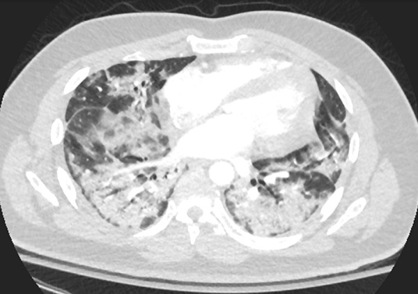
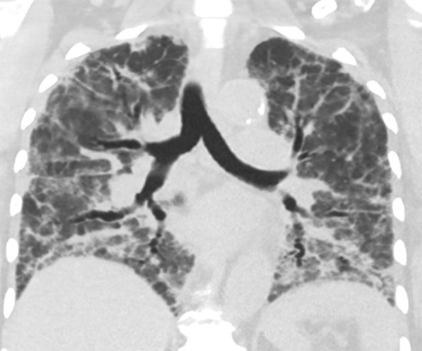
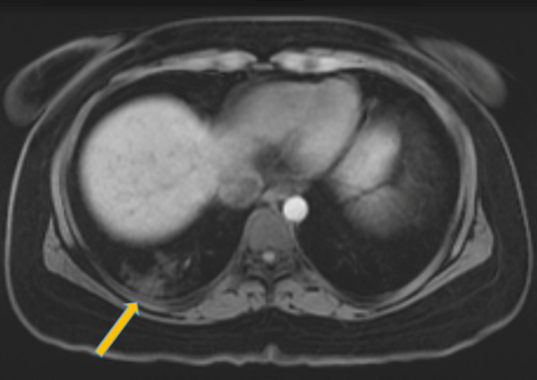
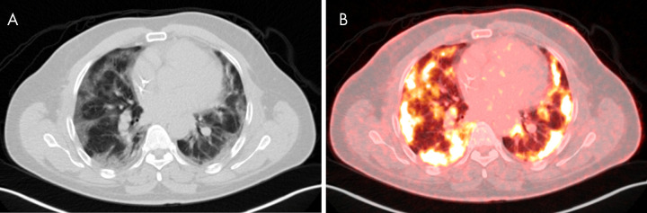
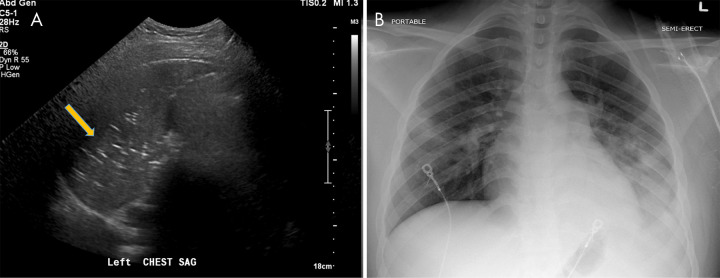
In this article we will review the imaging features of coronavirus disease 2019 (COVID-19) across multiple modalities, including radiography, CT, MRI, PET/CT, and US. Given that COVID-19 primarily affects the lung parenchyma by causing pneumonia, our directive is to focus on thoracic findings associated with COVID-19. We aim to enhance radiologists' understanding of this disease to help guide diagnosis and management. © RSNA, 2020.

摘要

在本文中,我们将回顾2019冠状病毒病(COVID-19)在多种影像学检查方式下的影像特征,包括X线摄影、CT、MRI、PET/CT和超声。鉴于COVID-19主要通过引起肺炎影响肺实质,我们的指导方针是重点关注与COVID-19相关的胸部表现。我们旨在加深放射科医生对这种疾病的理解,以帮助指导诊断和治疗。©RSNA,2020。

https://cdn.ncbi.nlm.nih.gov/pmc/blobs/a965/7977756/2e4e6313fc81/ryct.2020200210.fig12.jpg
https://cdn.ncbi.nlm.nih.gov/pmc/blobs/a965/7977756/30f136baec40/ryct.2020200210.fig1.jpg
https://cdn.ncbi.nlm.nih.gov/pmc/blobs/a965/7977756/6baf812545c3/ryct.2020200210.fig2.jpg
https://cdn.ncbi.nlm.nih.gov/pmc/blobs/a965/7977756/543534707b90/ryct.2020200210.fig3.jpg
https://cdn.ncbi.nlm.nih.gov/pmc/blobs/a965/7977756/c75544aa028d/ryct.2020200210.fig4.jpg
https://cdn.ncbi.nlm.nih.gov/pmc/blobs/a965/7977756/0ae373fa9397/ryct.2020200210.fig5.jpg
https://cdn.ncbi.nlm.nih.gov/pmc/blobs/a965/7977756/8957a13c4f26/ryct.2020200210.fig6.jpg
https://cdn.ncbi.nlm.nih.gov/pmc/blobs/a965/7977756/90f295ec84ed/ryct.2020200210.fig7.jpg
https://cdn.ncbi.nlm.nih.gov/pmc/blobs/a965/7977756/1ded43bb69b8/ryct.2020200210.fig8.jpg
https://cdn.ncbi.nlm.nih.gov/pmc/blobs/a965/7977756/c352ae09e96f/ryct.2020200210.fig9.jpg
https://cdn.ncbi.nlm.nih.gov/pmc/blobs/a965/7977756/f7355defa04f/ryct.2020200210.fig10.jpg
https://cdn.ncbi.nlm.nih.gov/pmc/blobs/a965/7977756/b2f83c2b583f/ryct.2020200210.fig11.jpg
https://cdn.ncbi.nlm.nih.gov/pmc/blobs/a965/7977756/2e4e6313fc81/ryct.2020200210.fig12.jpg

相似文献

1
COVID-19: A Multimodality Review of Radiologic Techniques, Clinical Utility, and Imaging Features.新型冠状病毒肺炎:放射学技术、临床应用及影像特征的多模态综述
Radiol Cardiothorac Imaging. 2020 Jun 1;2(3):e200210. doi: 10.1148/ryct.2020200210. eCollection 2020 Jun.
2
Artificial Intelligence Augmentation of Radiologist Performance in Distinguishing COVID-19 from Pneumonia of Other Origin at Chest CT.人工智能增强放射科医生在胸部 CT 上区分 COVID-19 与其他病因肺炎的性能。
Radiology. 2020 Sep;296(3):E156-E165. doi: 10.1148/radiol.2020201491. Epub 2020 Apr 27.
3
Performance of Radiologists in Differentiating COVID-19 from Non-COVID-19 Viral Pneumonia at Chest CT.放射科医生在胸部 CT 鉴别 COVID-19 与非 COVID-19 病毒性肺炎中的表现。
Radiology. 2020 Aug;296(2):E46-E54. doi: 10.1148/radiol.2020200823. Epub 2020 Mar 10.
4
Radiological Society of North America Expert Consensus Statement on Reporting Chest CT Findings Related to COVID-19. Endorsed by the Society of Thoracic Radiology, the American College of Radiology, and RSNA - Secondary Publication.北美放射学会关于报告与 COVID-19 相关的胸部 CT 结果的专家共识声明。得到胸放射学会、美国放射学会和 RSNA 的认可 - 二次出版物。
J Thorac Imaging. 2020 Jul;35(4):219-227. doi: 10.1097/RTI.0000000000000524.
5
Diagnosis of Coronavirus Disease 2019 Pneumonia by Using Chest Radiography: Value of Artificial Intelligence.利用胸部 X 线摄影诊断 2019 年冠状病毒病肺炎:人工智能的价值。
Radiology. 2021 Feb;298(2):E88-E97. doi: 10.1148/radiol.2020202944. Epub 2020 Sep 24.
6
RSNA Expert Consensus Statement on Reporting Chest CT Findings Related to COVID-19: Interobserver Agreement Between Chest Radiologists.RSNA 专家关于报告与 COVID-19 相关的胸部 CT 结果的共识声明:胸部放射科医生之间的观察者间一致性。
Can Assoc Radiol J. 2021 Feb;72(1):159-166. doi: 10.1177/0846537120938328. Epub 2020 Jul 2.
7
Role of Imaging in Diagnosis and Management of COVID-19: A Multiorgan Multimodality Imaging Review.影像学在新型冠状病毒肺炎诊断与管理中的作用:多器官多模态影像学综述
Front Med (Lausanne). 2021 Nov 8;8:765975. doi: 10.3389/fmed.2021.765975. eCollection 2021.
8
Pictorial Review of Pleural Disease: Multimodality Imaging and Differential Diagnosis.胸膜疾病的影像学表现:多模态成像与鉴别诊断。
Radiographics. 2024 Apr;44(4):e230079. doi: 10.1148/rg.230079.
9
Thoracic Imaging Findings of Multisystem Inflammatory Syndrome in Children Associated with COVID-19: What Radiologists Need to Know Now.与新型冠状病毒肺炎相关的儿童多系统炎症综合征的胸部影像学表现:放射科医生目前需要了解的内容
Radiol Cardiothorac Imaging. 2020 Jul 30;2(4):e200346. doi: 10.1148/ryct.2020200346. eCollection 2020 Aug.
10
Detection of Unsuspected Coronavirus Disease 2019 Cases by Computed Tomography and Retrospective Implementation of the Radiological Society of North America/Society of Thoracic Radiology/American College of Radiology Consensus Guidelines.利用计算机断层扫描检测未被怀疑的 2019 年冠状病毒病病例,并回顾性实施北美放射学会/胸放射学会/美国放射学院共识指南。
J Thorac Imaging. 2020 Nov 1;35(6):346-353. doi: 10.1097/RTI.0000000000000542.

引用本文的文献

1
Differentiation of COVID-19 from other types of viral pneumonia and severity scoring on baseline chest radiographs: Comparison of deep learning with multi-reader evaluation.基于胸部X光片将新型冠状病毒肺炎与其他类型病毒性肺炎进行鉴别及严重程度评分:深度学习与多位阅片者评估的比较
PLoS One. 2025 Jul 29;20(7):e0328061. doi: 10.1371/journal.pone.0328061. eCollection 2025.
2
The Use of the Modified Brixia Score for Predicting Mortality and Acute Respiratory Distress Syndrome in Patients with COVID-19 Pneumonia: What Have We Learned?改良布里夏评分在预测新型冠状病毒肺炎患者死亡率和急性呼吸窘迫综合征中的应用:我们学到了什么?
Diagnostics (Basel). 2025 Jun 1;15(11):1409. doi: 10.3390/diagnostics15111409.
3

本文引用的文献

1
Serial Quantitative Chest CT Assessment of COVID-19: A Deep Learning Approach.COVID-19的胸部CT序列定量评估:一种深度学习方法。
Radiol Cardiothorac Imaging. 2020 Mar 30;2(2):e200075. doi: 10.1148/ryct.2020200075. eCollection 2020 Apr.
2
Imaging Profile of the COVID-19 Infection: Radiologic Findings and Literature Review.新型冠状病毒肺炎感染的影像学表现:放射学发现与文献综述
Radiol Cardiothorac Imaging. 2020 Feb 13;2(1):e200034. doi: 10.1148/ryct.2020200034. eCollection 2020 Feb.
3
Deep Learning Enables Accurate Diagnosis of Novel Coronavirus (COVID-19) With CT Images.
Clinical implications of teleradiology in rheumatic and musculoskeletal diseases: improving rheumatic care.
远程放射学在风湿性和肌肉骨骼疾病中的临床意义:改善风湿性疾病护理
Rheumatol Int. 2025 Feb 13;45(3):51. doi: 10.1007/s00296-025-05810-w.
4
Development and evaluation of an F-labeled nanobody to target SARS-CoV-2's spike protein.一种靶向严重急性呼吸综合征冠状病毒2(SARS-CoV-2)刺突蛋白的F标记纳米抗体的研发与评估。
Front Nucl Med. 2022 Nov 23;2:1033697. doi: 10.3389/fnume.2022.1033697. eCollection 2022.
5
A review of current effective COVID-19 testing methods and quality control.当前有效 COVID-19 检测方法及质量控制综述。
Arch Microbiol. 2023 May 17;205(6):239. doi: 10.1007/s00203-023-03579-9.
6
Multi-head deep learning framework for pulmonary disease detection and severity scoring with modified progressive learning.用于肺部疾病检测和严重程度评分的多头深度学习框架及改进的渐进式学习
Biomed Signal Process Control. 2023 Aug;85:104855. doi: 10.1016/j.bspc.2023.104855. Epub 2023 Mar 24.
7
Pneumomediastinum and Pneumopericardium as Uncommon Complications of COVID-19 Infection: A Review Article.纵隔气肿和心包积气作为新型冠状病毒肺炎感染的罕见并发症:一篇综述文章
Cureus. 2022 Oct 13;14(10):e30244. doi: 10.7759/cureus.30244. eCollection 2022 Oct.
8
Pembrolizumab-related pneumonitis in a patient with COVID-19 infection.一名新冠病毒感染患者发生的帕博利珠单抗相关性肺炎。
Singapore Med J. 2023 Jul;64(7):454-458. doi: 10.11622/smedj.2022083.
9
Review of Thoracic Imaging Manifestations of COVID-19 and Other Pathologic Coronaviruses.COVID-19 及其他病理性冠状病毒的胸部影像学表现综述。
Radiol Clin North Am. 2022 May;60(3):359-369. doi: 10.1016/j.rcl.2022.01.004. Epub 2022 Jan 11.
10
Multimodal covid network: Multimodal bespoke convolutional neural network architectures for COVID-19 detection from chest X-ray's and computerized tomography scans.多模态新冠网络:用于从胸部X光和计算机断层扫描中检测新冠病毒的多模态定制卷积神经网络架构。
Int J Imaging Syst Technol. 2022 May;32(3):704-716. doi: 10.1002/ima.22712. Epub 2022 Jan 31.
深度学习利用 CT 图像准确诊断新型冠状病毒(COVID-19)。
IEEE/ACM Trans Comput Biol Bioinform. 2021 Nov-Dec;18(6):2775-2780. doi: 10.1109/TCBB.2021.3065361. Epub 2021 Dec 8.
4
A deep learning algorithm using CT images to screen for Corona virus disease (COVID-19).利用 CT 图像进行冠状病毒病(COVID-19)筛查的深度学习算法。
Eur Radiol. 2021 Aug;31(8):6096-6104. doi: 10.1007/s00330-021-07715-1. Epub 2021 Feb 24.
5
Acute cerebrovascular disease following COVID-19: a single center, retrospective, observational study.新型冠状病毒肺炎后急性脑血管病:一项单中心、回顾性、观察性研究。
Stroke Vasc Neurol. 2020 Sep;5(3):279-284. doi: 10.1136/svn-2020-000431. Epub 2020 Jul 2.
6
Evaluation of variation in D-dimer levels among COVID-19 and bacterial pneumonia: a retrospective analysis.评估 COVID-19 和细菌性肺炎患者 D-二聚体水平的变化:一项回顾性分析。
J Thromb Thrombolysis. 2020 Oct;50(3):548-557. doi: 10.1007/s11239-020-02171-y.
7
SARS-CoV-2 Gastrointestinal Infection Causing Hemorrhagic Colitis: Implications for Detection and Transmission of COVID-19 Disease.严重急性呼吸综合征冠状病毒2型(SARS-CoV-2)胃肠道感染引发出血性结肠炎:对2019冠状病毒病(COVID-19)检测与传播的影响
Am J Gastroenterol. 2020 Jun;115(6):942-946. doi: 10.14309/ajg.0000000000000667.
8
Large-Vessel Stroke as a Presenting Feature of Covid-19 in the Young.大血管卒中作为年轻人新冠病毒病的首发特征
N Engl J Med. 2020 May 14;382(20):e60. doi: 10.1056/NEJMc2009787. Epub 2020 Apr 28.
9
ISTH interim guidance on recognition and management of coagulopathy in COVID-19.国际血栓与止血学会(ISTH)关于新冠病毒病(COVID-19)凝血病识别与管理的临时指南。
J Thromb Haemost. 2020 May;18(5):1023-1026. doi: 10.1111/jth.14810. Epub 2020 Apr 27.
10
Pulmonary Embolism in Patients With COVID-19: Awareness of an Increased Prevalence.2019冠状病毒病患者的肺栓塞:对患病率增加的认识
Circulation. 2020 Jul 14;142(2):184-186. doi: 10.1161/CIRCULATIONAHA.120.047430. Epub 2020 Apr 24.