Bonardel Gerald, Dupont Axel, Decazes Pierre, Queneau Mathieu, Modzelewski Romain, Coulot Jeremy, Le Calvez Nicolas, Hapdey Sébastien
Nuclear Medicine, Centre Cardiologique du Nord, Saint-Denis, France.
Nuclear Medicine, Hopital Delafontaine, Saint-Denis, France.
EJNMMI Phys. 2022 May 11;9(1):36. doi: 10.1186/s40658-022-00465-z.
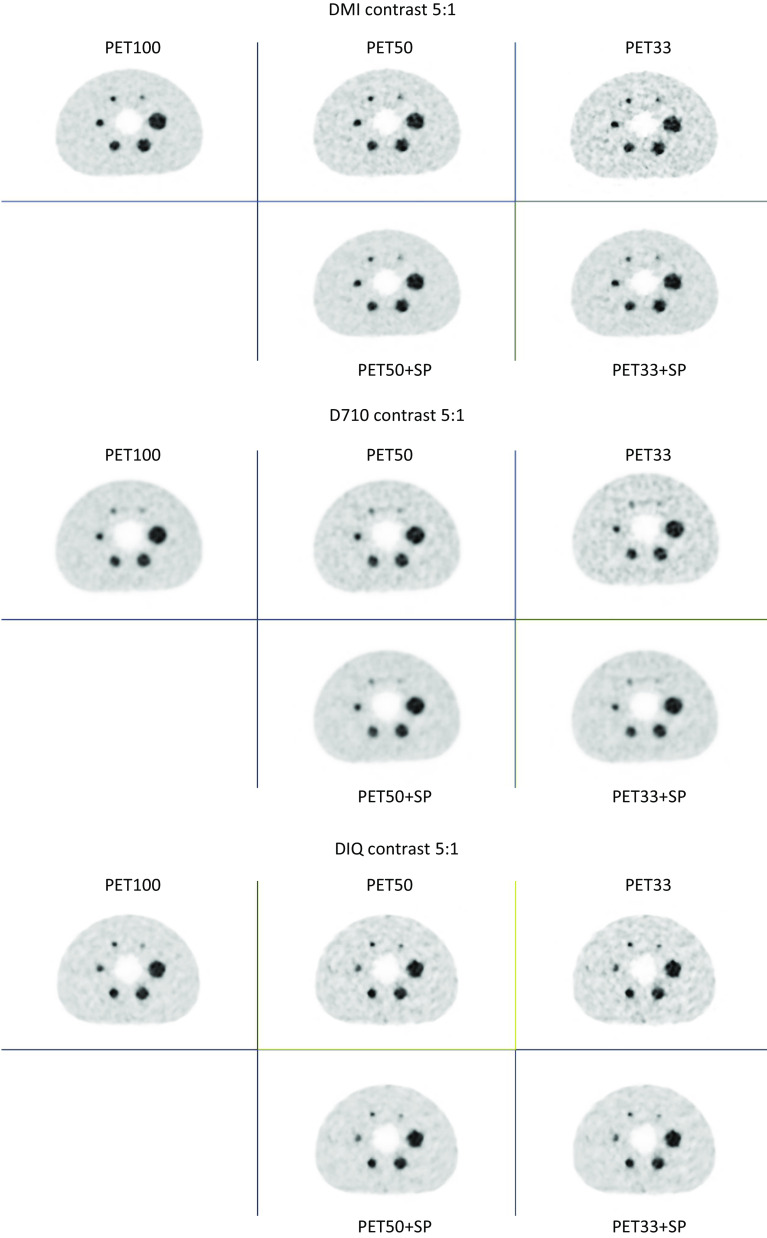
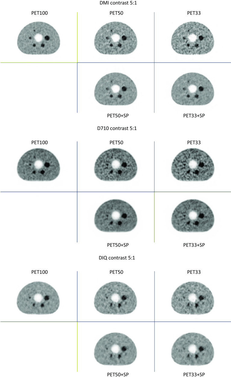
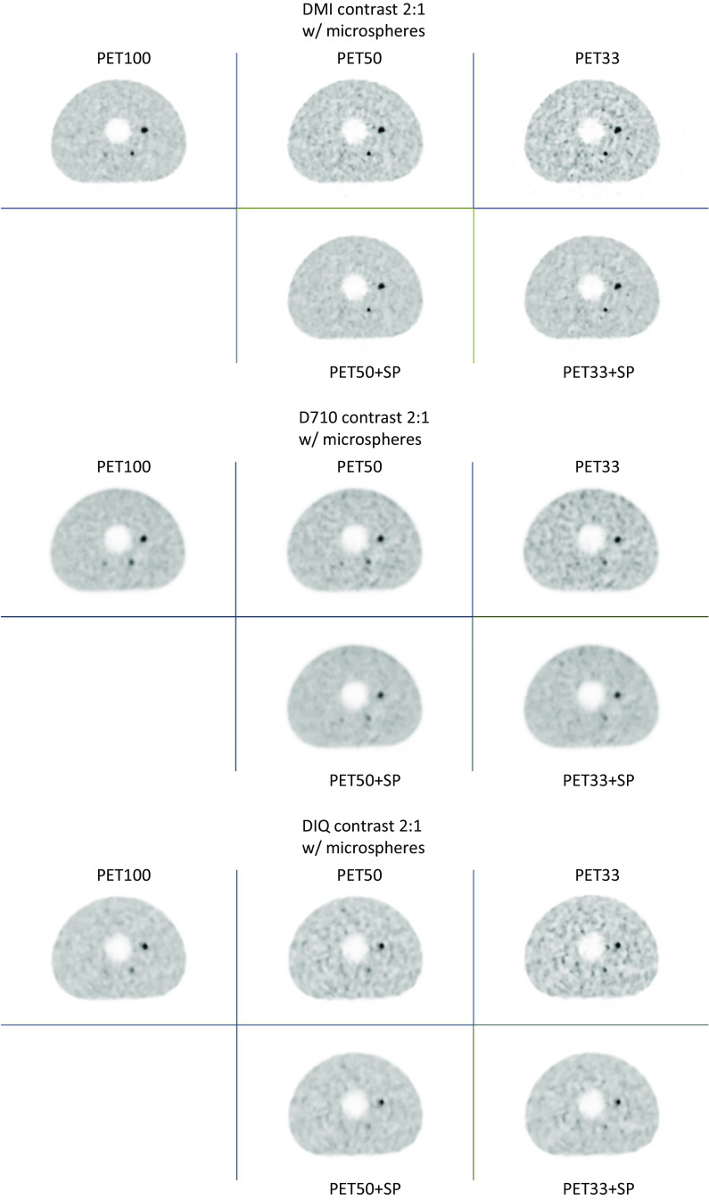
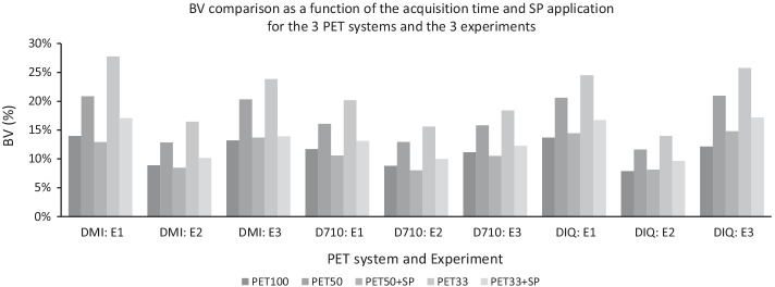
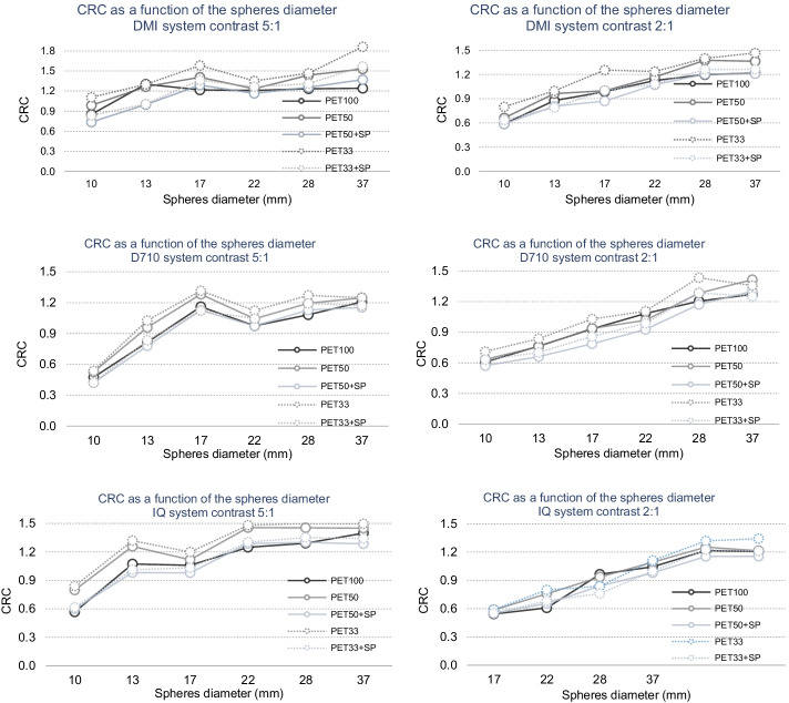
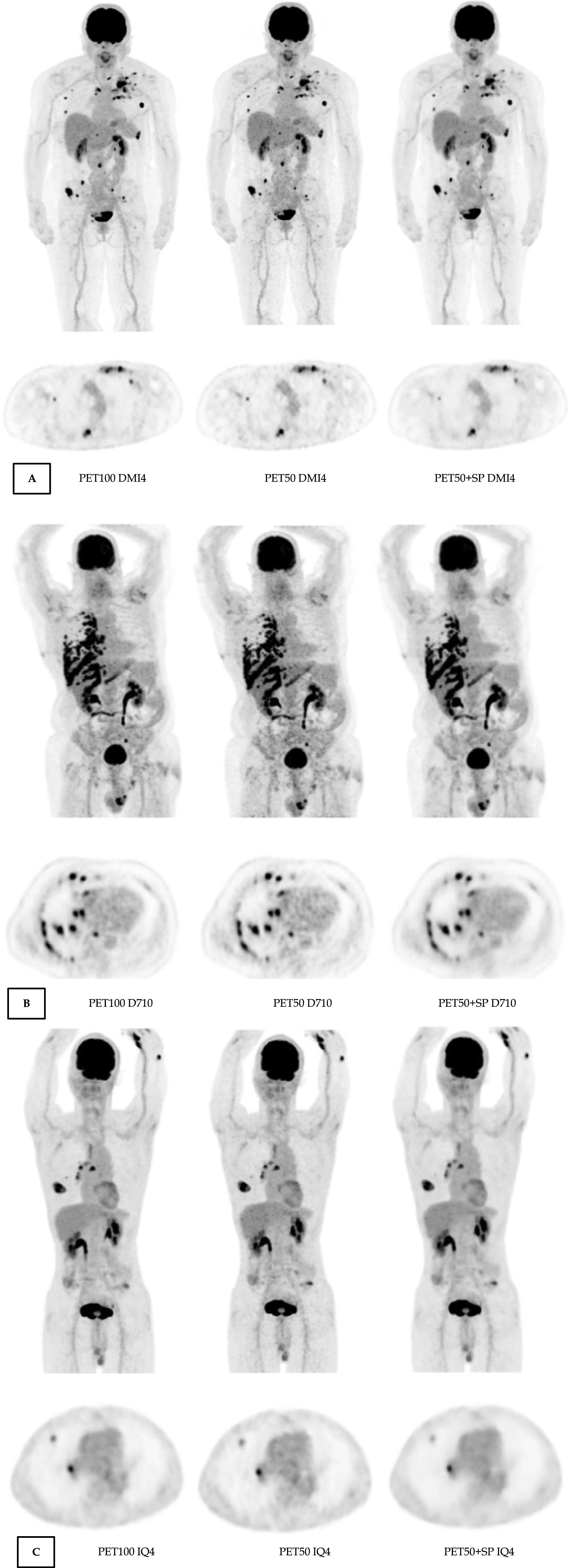
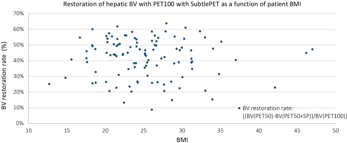
PET/CT image quality is directly influenced by the F-18-FDG injected activity. The higher the injected activity, the less noise in the reconstructed images but the more radioactive staff exposition. A new FDA cleared software has been introduced to obtain clinical PET images, acquired at 25% of the count statistics considering US practices. Our aim is to determine the limits of a deep learning based denoising algorithm (SubtlePET) applied to statistically reduced PET raw data from 3 different last generation PET scanners in comparison to the regular acquisition in phantom and patients, considering the European guidelines for radiotracer injection activities. Images of low and high contrasted (SBR = 2 and 5) spheres of the IEC phantom and high contrast (SBR = 5) of micro-spheres of Jaszczak phantom were acquired on 3 different PET devices. 110 patients with different pathologies were included. The data was acquired in list-mode and retrospectively reconstructed with the regular acquisition count statistic (PET100), 50% reduction in counts (PET50) and 66% reduction in counts (PET33). These count reduced images were post-processed with SubtlePET to obtain PET50 + SP and PET33 + SP images. Patient image quality was scored by 2 senior nuclear physicians. Peak-signal-to-Noise and Structural similarity metrics were computed to compare the low count images to regular acquisition (PET100).
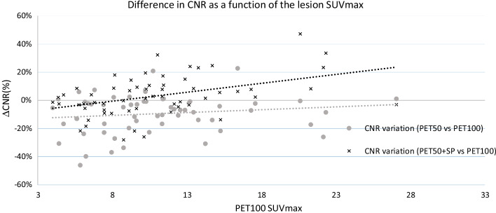
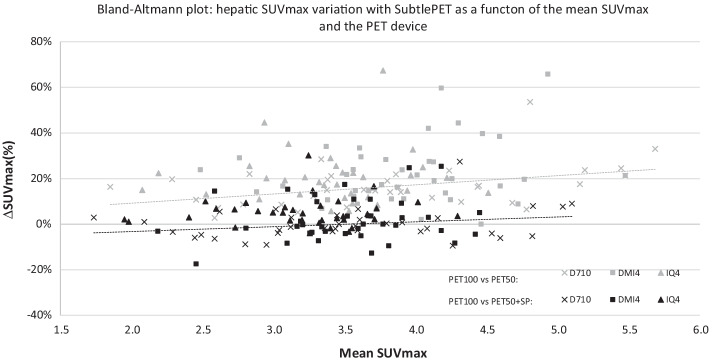
SubtlePET reliably denoised the images and maintained the SUV values in PET50 + SP. SubtlePET enhanced images (PET33 + SP) had slightly increased noise compared to PET100 and could lead to a potential loss of information in terms of lesion detectability. Regarding the patient datasets, the PET100 and PET50 + SP were qualitatively comparable. The SubtlePET algorithm was able to correctly recover the SUV values of the lesions and maintain a noise level equivalent to full-time images.
Based on our results, SubtlePET is adapted in clinical practice for half-time or half-dose acquisitions based on European recommended injected dose of 3 MBq/kg without diagnostic confidence loss.
PET/CT图像质量直接受F-18-FDG注射活度影响。注射活度越高,重建图像中的噪声越少,但工作人员受到的放射性暴露越多。一种新的获得美国食品药品监督管理局(FDA)批准的软件已被引入,用于获取临床PET图像,该图像是按照美国的做法在计数统计的25%时采集的。我们的目的是根据放射性示踪剂注射活度的欧洲指南,确定一种基于深度学习的去噪算法(SubtlePET)应用于来自3种不同的新一代PET扫描仪的统计减少的PET原始数据时的局限性,与在体模和患者中的常规采集进行比较。在3种不同的PET设备上采集了国际电工委员会(IEC)体模的低对比度和高对比度(SBR = 2和5)球体以及Jaszczak体模的高对比度(SBR = 5)微球体的图像。纳入了110例患有不同疾病的患者。数据以列表模式采集,并使用常规采集计数统计(PET100)、计数减少50%(PET50)和计数减少66%(PET33)进行回顾性重建。这些计数减少的图像用SubtlePET进行后处理,以获得PET50 + SP和PET33 + SP图像。由2名资深核医学医师对患者图像质量进行评分。计算峰值信噪比和结构相似性指标,以将低计数图像与常规采集(PET100)进行比较。
SubtlePET可靠地对图像进行了去噪,并在PET50 + SP中保持了SUV值(标准化摄取值)。与PET100相比,SubtlePET增强图像(PET33 + SP)的噪声略有增加,并且在病变可检测性方面可能导致信息潜在丢失。关于患者数据集,PET100和PET50 + SP在质量上具有可比性。SubtlePET算法能够正确恢复病变的SUV值,并保持与全时图像相当的噪声水平。
基于我们的结果,根据欧洲推荐的3 MBq/kg注射剂量,SubtlePET适用于临床实践中的半时或半剂量采集,且不会损失诊断置信度。