• 文献检索
  • 文档翻译
  • 深度研究
  • 学术资讯
  • Suppr Zotero 插件Zotero 插件
  • 邀请有礼
  • 套餐&价格
  • 历史记录
应用&插件
Suppr Zotero 插件Zotero 插件浏览器插件Mac 客户端Windows 客户端微信小程序
定价
高级版会员购买积分包购买API积分包
服务
文献检索文档翻译深度研究API 文档MCP 服务
关于我们
关于 Suppr公司介绍联系我们用户协议隐私条款
关注我们

Suppr 超能文献

核心技术专利:CN118964589B侵权必究
粤ICP备2023148730 号-1Suppr @ 2026

文献检索

告别复杂PubMed语法,用中文像聊天一样搜索,搜遍4000万医学文献。AI智能推荐,让科研检索更轻松。

立即免费搜索

文件翻译

保留排版,准确专业,支持PDF/Word/PPT等文件格式,支持 12+语言互译。

免费翻译文档

深度研究

AI帮你快速写综述,25分钟生成高质量综述,智能提取关键信息,辅助科研写作。

立即免费体验

碳-11治疗设施技术设计报告

Technical Design Report for a Carbon-11 Treatment Facility.

作者信息

Penescu Liviu, Stora Thierry, Stegemann Simon, Pitters Johanna, Fiorina Elisa, Augusto Ricardo Dos Santos, Schmitzer Claus, Wenander Fredrik, Parodi Katia, Ferrari Alfredo, Cocolios Thomas E

机构信息

Abstract Landscapes, Montpellier, France.

European Organization for Nuclear Research (CERN), Geneva, Switzerland.

出版信息

Front Med (Lausanne). 2022 Apr 25;8:697235. doi: 10.3389/fmed.2021.697235. eCollection 2021.

DOI:10.3389/fmed.2021.697235
PMID:35547661
原文链接:https://pmc.ncbi.nlm.nih.gov/articles/PMC9081534/
Abstract

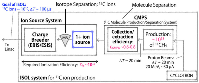
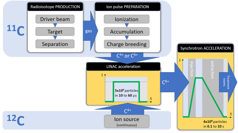
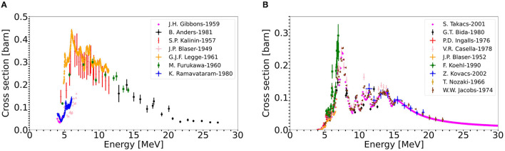
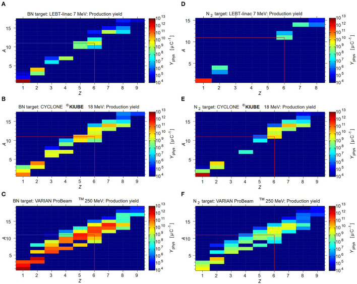
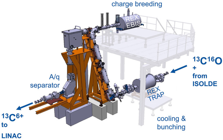
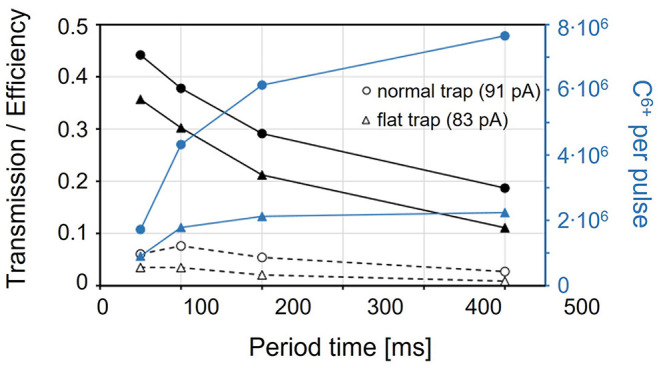
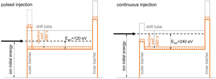
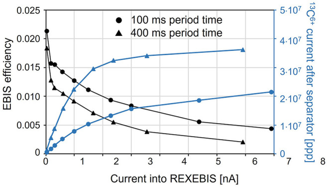
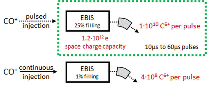
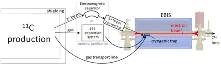
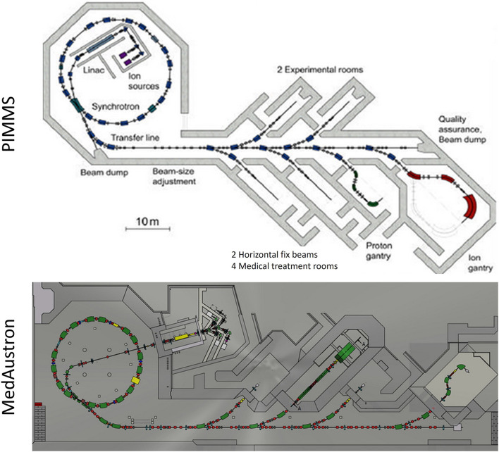
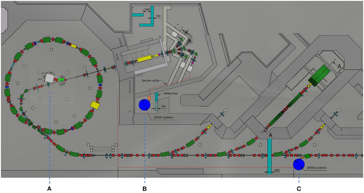
Particle therapy relies on the advantageous dose deposition which permits to highly conform the dose to the target and better spare the surrounding healthy tissues and organs at risk with respect to conventional radiotherapy. In the case of treatments with heavier ions (like carbon ions already clinically used), another advantage is the enhanced radiobiological effectiveness due to high linear energy transfer radiation. These particle therapy advantages are unfortunately not thoroughly exploited due to particle range uncertainties. The possibility to monitor the compliance between the ongoing and prescribed dose distribution is a crucial step toward new optimizations in treatment planning and adaptive therapy. The Positron Emission Tomography (PET) is an established quantitative 3D imaging technique for particle treatment verification and, among the isotopes used for PET imaging, the C has gained more attention from the scientific and clinical communities for its application as new radioactive projectile for particle therapy. This is an interesting option clinically because of an enhanced imaging potential, without dosimetry drawbacks; technically, because the stable isotope C is successfully already in use in clinics. The MEDICIS-Promed network led an initiative to study the possible technical solutions for the implementation of C radioisotopes in an accelerator-based particle therapy center. We present here the result of this study, consisting in a Technical Design Report for a C Treatment Facility. The clinical usefulness is reviewed based on existing experimental data, complemented by Monte Carlo simulations using the FLUKA code. The technical analysis starts from reviewing the layout and results of the facilities which produced C beams in the past, for testing purposes. It then focuses on the elaboration of the feasible upgrades of an existing C particle therapy center, to accommodate the production of C beams for therapy. The analysis covers the options to produce the C atoms in sufficient amounts (as required for therapy), to ionize them as required by the existing accelerator layouts, to accelerate and transport them to the irradiation rooms. The results of the analysis and the identified challenges define the possible implementation scenario and timeline.

摘要

粒子治疗依赖于有利的剂量沉积,这使得能够高度精确地将剂量适形于靶区,并相对于传统放疗更好地保护周围健康组织和危及器官。在使用较重离子(如已临床应用的碳离子)进行治疗的情况下,另一个优势是由于高线性能量传递辐射而增强的放射生物学效应。不幸的是,由于粒子射程的不确定性,这些粒子治疗的优势并未得到充分利用。监测正在进行的剂量分布与规定剂量分布之间的一致性,是朝着治疗计划和自适应治疗的新优化迈出的关键一步。正电子发射断层扫描(PET)是一种成熟的用于粒子治疗验证的定量三维成像技术,在用于PET成像的同位素中,碳-11因其作为粒子治疗新的放射性射弹的应用而受到科学界和临床界更多的关注。这在临床上是一个有趣的选择,因为其成像潜力增强,且没有剂量测定方面的缺点;从技术上讲,是因为稳定同位素碳-12已成功应用于临床。MEDICIS-Promed网络发起了一项倡议,研究在基于加速器的粒子治疗中心实施碳-11放射性同位素的可能技术方案。我们在此展示这项研究的结果,即一份碳-11治疗设施的技术设计报告。基于现有实验数据对临床实用性进行了评估,并辅以使用FLUKA代码的蒙特卡罗模拟。技术分析首先回顾过去为测试目的而产生碳-11束流的设施的布局和结果。然后重点阐述对现有碳-12粒子治疗中心进行可行升级的方案,以适应治疗用碳-11束流的生产。分析涵盖了以足够数量(治疗所需)生产碳-11原子、按照现有加速器布局的要求将其电离、加速并输送至照射室的各种选项。分析结果和所确定的挑战确定了可能的实施方案和时间表。

https://cdn.ncbi.nlm.nih.gov/pmc/blobs/69c2/9081534/b986b6f35f32/fmed-08-697235-g0015.jpg
https://cdn.ncbi.nlm.nih.gov/pmc/blobs/69c2/9081534/19d20331afff/fmed-08-697235-g0001.jpg
https://cdn.ncbi.nlm.nih.gov/pmc/blobs/69c2/9081534/9daa58ae7097/fmed-08-697235-g0002.jpg
https://cdn.ncbi.nlm.nih.gov/pmc/blobs/69c2/9081534/895772d346ec/fmed-08-697235-g0003.jpg
https://cdn.ncbi.nlm.nih.gov/pmc/blobs/69c2/9081534/2ab5a130e88e/fmed-08-697235-g0004.jpg
https://cdn.ncbi.nlm.nih.gov/pmc/blobs/69c2/9081534/6354cea2bd56/fmed-08-697235-g0005.jpg
https://cdn.ncbi.nlm.nih.gov/pmc/blobs/69c2/9081534/3b63df1f558d/fmed-08-697235-g0006.jpg
https://cdn.ncbi.nlm.nih.gov/pmc/blobs/69c2/9081534/866552e83e00/fmed-08-697235-g0007.jpg
https://cdn.ncbi.nlm.nih.gov/pmc/blobs/69c2/9081534/3a407ed6927a/fmed-08-697235-g0008.jpg
https://cdn.ncbi.nlm.nih.gov/pmc/blobs/69c2/9081534/93ce695e1c43/fmed-08-697235-g0009.jpg
https://cdn.ncbi.nlm.nih.gov/pmc/blobs/69c2/9081534/ad32b9989d2d/fmed-08-697235-g0010.jpg
https://cdn.ncbi.nlm.nih.gov/pmc/blobs/69c2/9081534/aea2b59e214c/fmed-08-697235-g0011.jpg
https://cdn.ncbi.nlm.nih.gov/pmc/blobs/69c2/9081534/c2b229696af4/fmed-08-697235-g0012.jpg
https://cdn.ncbi.nlm.nih.gov/pmc/blobs/69c2/9081534/0a68fcc10211/fmed-08-697235-g0013.jpg
https://cdn.ncbi.nlm.nih.gov/pmc/blobs/69c2/9081534/a8d1d175931b/fmed-08-697235-g0014.jpg
https://cdn.ncbi.nlm.nih.gov/pmc/blobs/69c2/9081534/b986b6f35f32/fmed-08-697235-g0015.jpg

相似文献

1
Technical Design Report for a Carbon-11 Treatment Facility.碳-11治疗设施技术设计报告
Front Med (Lausanne). 2022 Apr 25;8:697235. doi: 10.3389/fmed.2021.697235. eCollection 2021.
2
Experimental validation of the FLUKA Monte Carlo code for dose and [Formula: see text]-emitter predictions of radioactive ion beams.放射性离子束剂量和 [Formula: see text]-发射体预测的 FLUKA 蒙特卡罗代码的实验验证。
Phys Med Biol. 2018 Oct 26;63(21):215014. doi: 10.1088/1361-6560/aae431.
3
Depth dose measurements in water for C and C beams with therapy relevant energies.在水中对具有治疗相关能量的碳离子束和碳离子束进行深度剂量测量。
Nucl Instrum Methods Phys Res A. 2022 Nov 11;1043:167464. doi: 10.1016/j.nima.2022.167464. Epub 2022 Oct 1.
4
Investigating the accuracy of the FLUKA code for transport of therapeutic ion beams in matter.研究用于治疗性离子束在物质中传输的FLUKA代码的准确性。
Phys Med Biol. 2006 Sep 7;51(17):4385-98. doi: 10.1088/0031-9155/51/17/017. Epub 2006 Aug 15.
5
In-beam PET monitoring of mono-energetic (16)O and (12)C beams: experiments and FLUKA simulations for homogeneous targets.单能(16)O和(12)C束流的束内正电子发射断层扫描监测:均匀靶体的实验与FLUKA模拟
Phys Med Biol. 2009 Jul 7;54(13):3979-96. doi: 10.1088/0031-9155/54/13/003. Epub 2009 Jun 3.
6
Experimental investigation of the characteristics of radioactive beams for heavy ion therapy.重离子治疗中放射性束流特性的实验研究。
Med Phys. 2020 Jul;47(7):3123-3132. doi: 10.1002/mp.14177. Epub 2020 May 11.
7
Radioactive Beams in Particle Therapy: Past, Present, and Future.粒子治疗中的放射性束流:过去、现在与未来。
Front Phys. 2020 Aug 28;8:00326. doi: 10.3389/fphy.2020.00326.
8
Precision of the PET activity range during irradiation withC,C, andC beams.利用C、C 和 C 射线进行放射治疗时的 PET 活性范围精度。
Phys Med Biol. 2022 Dec 19;68(1). doi: 10.1088/1361-6560/aca5e8.
9
Range verification of radioactive ion beams of C and O using in-beam PET imaging.使用放射性离子束内在线正电子发射断层成像技术对 C 和 O 进行放射性离子束射程验证。
Phys Med Biol. 2019 Jul 16;64(14):145014. doi: 10.1088/1361-6560/ab25ce.
10
Secondary radiation measurements for particle therapy applications: prompt photons produced by He, C and O ion beams in a PMMA target.用于粒子治疗应用的二次辐射测量:氦、碳和氧离子束在聚甲基丙烯酸甲酯靶中产生的瞬发光子。
Phys Med Biol. 2017 Feb 21;62(4):1438-1455. doi: 10.1088/1361-6560/62/4/1438. Epub 2017 Jan 23.

引用本文的文献

1
Quasi-real-time range monitoring by in-beam PET: a case for O.通过束流中PET进行的准实时范围监测:以O为例
Sci Rep. 2023 Nov 1;13(1):18788. doi: 10.1038/s41598-023-45122-2.
2
Radioactive Beams in Particle Therapy: Past, Present, and Future.粒子治疗中的放射性束流:过去、现在与未来。
Front Phys. 2020 Aug 28;8:00326. doi: 10.3389/fphy.2020.00326.

本文引用的文献

1
Radioactive Beams for Image-Guided Particle Therapy: The BARB Experiment at GSI.用于图像引导粒子治疗的放射性束流:GSI的BARB实验
Front Oncol. 2021 Aug 19;11:737050. doi: 10.3389/fonc.2021.737050. eCollection 2021.
2
Optimization of synchrotron based ion beam therapy facilities for treatment time reduction, options and the MedAustron development roadmap.基于同步加速器的离子束治疗设施的优化以减少治疗时间、选择和 MedAustron 发展路线图。
Phys Med. 2021 Jan;81:264-272. doi: 10.1016/j.ejmp.2020.11.029. Epub 2020 Dec 24.
3
Radioactive Beams in Particle Therapy: Past, Present, and Future.
粒子治疗中的放射性束流:过去、现在与未来。
Front Phys. 2020 Aug 28;8:00326. doi: 10.3389/fphy.2020.00326.
4
A filtering approach for PET and PG predictions in a proton treatment planning system.一种质子治疗计划系统中 PET 和 PG 预测的过滤方法。
Phys Med Biol. 2020 May 11;65(9):095014. doi: 10.1088/1361-6560/ab8146.
5
Monte Carlo investigation of the characteristics of radioactive beams for heavy ion therapy.蒙特卡罗方法研究重离子治疗用放射性束的特性。
Sci Rep. 2019 Apr 25;9(1):6537. doi: 10.1038/s41598-019-43073-1.
6
Including anatomical variations in robust optimization for head and neck proton therapy can reduce the need of adaptation.在头颈部质子治疗的稳健优化中纳入解剖学变异,可以减少适应的需要。
Radiother Oncol. 2019 Feb;131:127-134. doi: 10.1016/j.radonc.2018.12.008. Epub 2018 Dec 31.
7
FLUKA particle therapy tool for Monte Carlo independent calculation of scanned proton and carbon ion beam therapy.FLUKA 粒子治疗工具,用于独立计算扫描质子和碳离子束治疗的蒙特卡罗。
Phys Med Biol. 2019 Mar 29;64(7):075012. doi: 10.1088/1361-6560/ab02cb.
8
Experimental validation of the FLUKA Monte Carlo code for dose and [Formula: see text]-emitter predictions of radioactive ion beams.放射性离子束剂量和 [Formula: see text]-发射体预测的 FLUKA 蒙特卡罗代码的实验验证。
Phys Med Biol. 2018 Oct 26;63(21):215014. doi: 10.1088/1361-6560/aae431.
9
An overview of recent developments in FLUKA PET tools.FLUKA PET 工具近期发展概述。
Phys Med. 2018 Oct;54:189-199. doi: 10.1016/j.ejmp.2018.06.636. Epub 2018 Jul 2.
10
Monte Carlo simulation tool for online treatment monitoring in hadrontherapy with in-beam PET: A patient study.用于质子治疗在线治疗监测的蒙特卡罗模拟工具:一项患者研究。
Phys Med. 2018 Jul;51:71-80. doi: 10.1016/j.ejmp.2018.05.002. Epub 2018 May 7.