• 文献检索
  • 文档翻译
  • 深度研究
  • 学术资讯
  • Suppr Zotero 插件Zotero 插件
  • 邀请有礼
  • 套餐&价格
  • 历史记录
应用&插件
Suppr Zotero 插件Zotero 插件浏览器插件Mac 客户端Windows 客户端微信小程序
定价
高级版会员购买积分包购买API积分包
服务
文献检索文档翻译深度研究API 文档MCP 服务
关于我们
关于 Suppr公司介绍联系我们用户协议隐私条款
关注我们

Suppr 超能文献

核心技术专利:CN118964589B侵权必究
粤ICP备2023148730 号-1Suppr @ 2026

文献检索

告别复杂PubMed语法,用中文像聊天一样搜索,搜遍4000万医学文献。AI智能推荐,让科研检索更轻松。

立即免费搜索

文件翻译

保留排版,准确专业,支持PDF/Word/PPT等文件格式,支持 12+语言互译。

免费翻译文档

深度研究

AI帮你快速写综述,25分钟生成高质量综述,智能提取关键信息,辅助科研写作。

立即免费体验

视网膜血管炎综合更新:病因、表现及治疗

A Comprehensive Update on Retinal Vasculitis: Etiologies, Manifestations and Treatments.

作者信息

Agarwal Aniruddha, Rübsam Anne, Zur Bonsen Lynn, Pichi Francesco, Neri Piergiorgio, Pleyer Uwe

机构信息

The Eye Institute, Cleveland Clinic Abu Dhabi, Abu Dhabi P.O. Box 124140, United Arab Emirates.

Cleveland Clinic Lerner College of Medicine, Cleveland, OH 44195, USA.

出版信息

J Clin Med. 2022 Apr 30;11(9):2525. doi: 10.3390/jcm11092525.

DOI:10.3390/jcm11092525
PMID:35566655
原文链接:https://pmc.ncbi.nlm.nih.gov/articles/PMC9101900/
Abstract

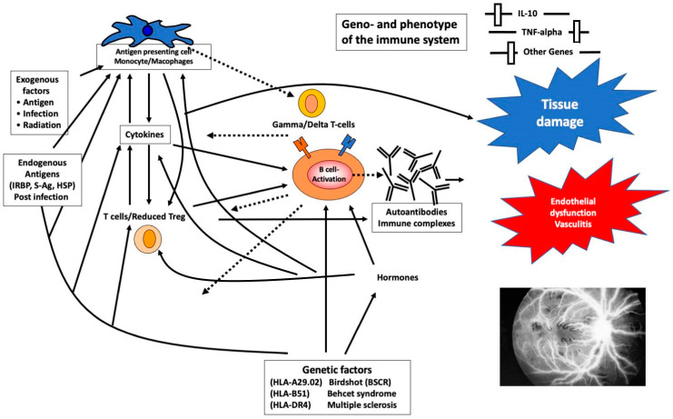
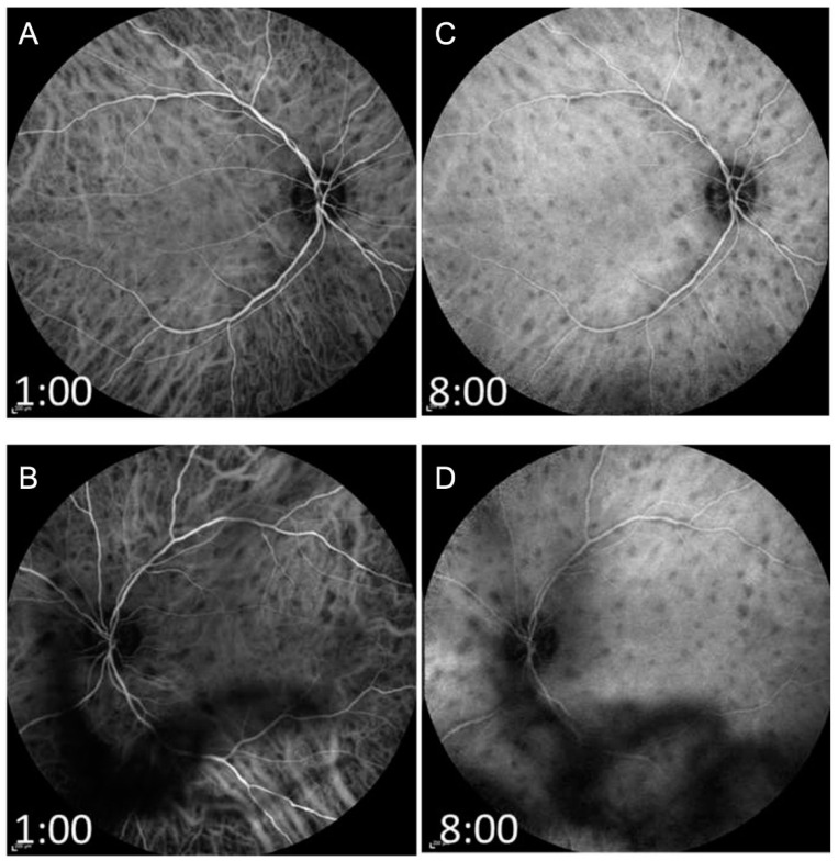
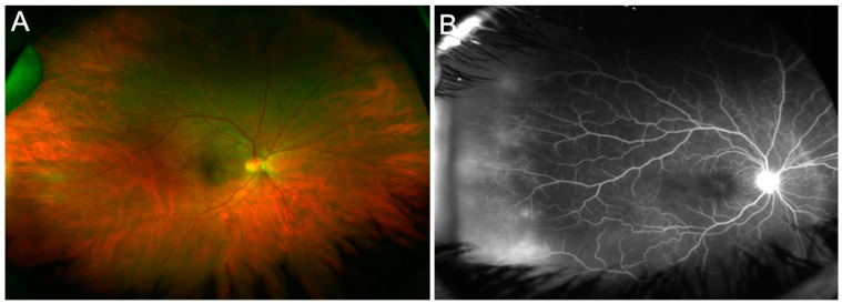
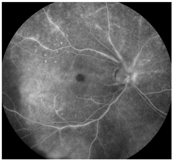
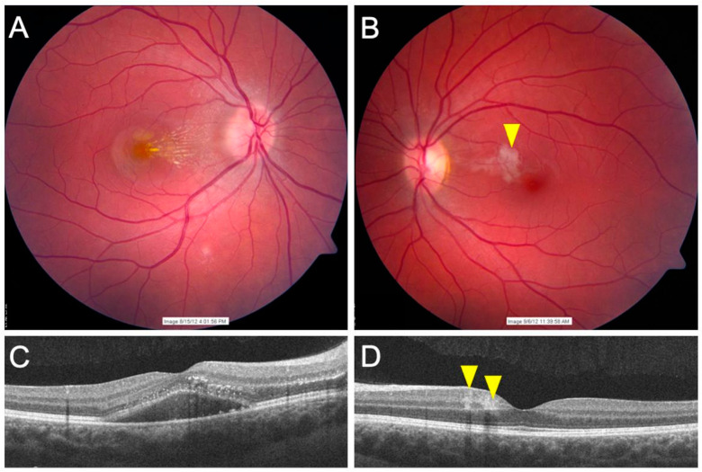
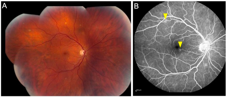
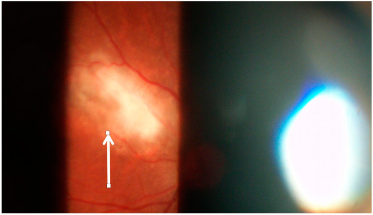
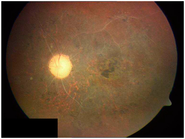
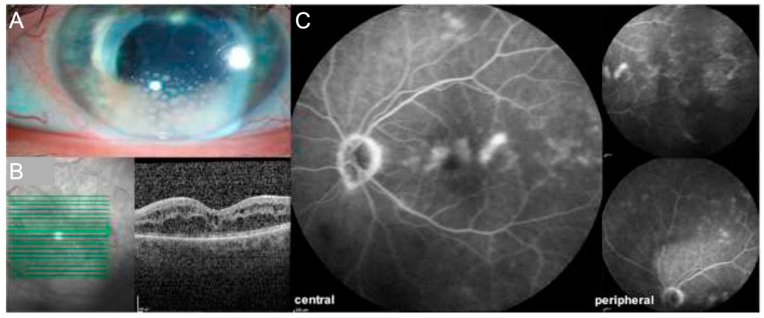
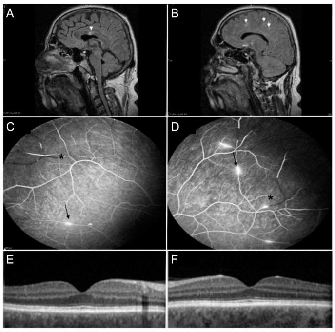
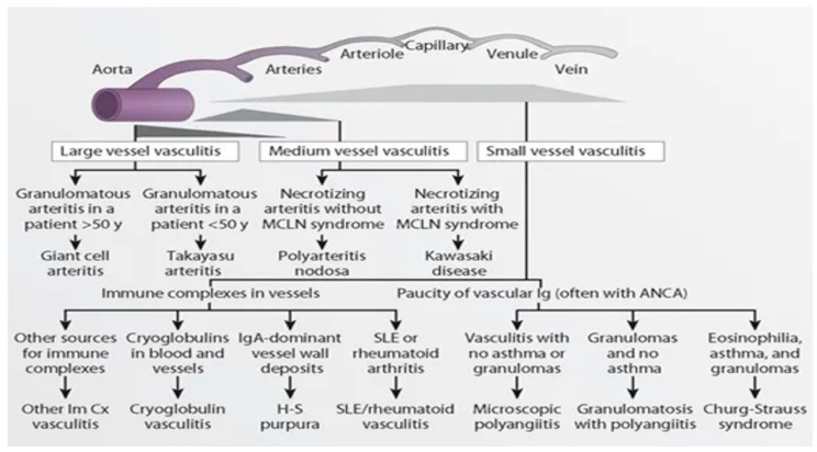
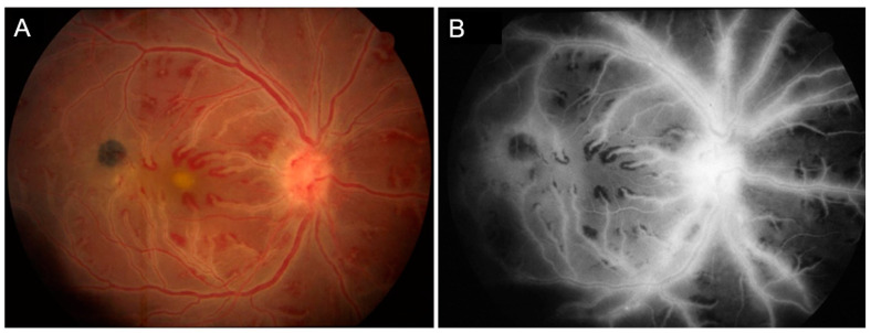
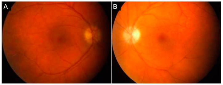
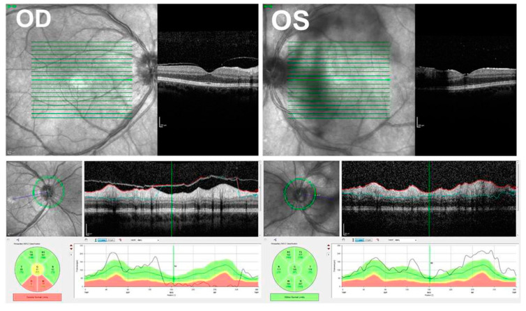
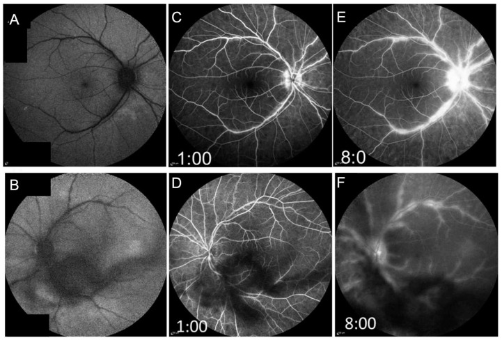
Retinal vasculitis is characterized by inflammatory involvement of retinal arterioles, venules and/or capillaries and can be associated with a myriad of systemic and ophthalmic diseases. In this review, we have comprehensively discussed the etiologies, clinical manifestations, and presentations of retinal vasculitis. We have also included newer advances in imaging in retinal vasculitis such as OCTA and widefield imaging.

摘要

视网膜血管炎的特征是视网膜小动脉、小静脉和/或毛细血管发生炎症,并且可能与多种全身和眼科疾病相关。在本综述中,我们全面讨论了视网膜血管炎的病因、临床表现和症状。我们还纳入了视网膜血管炎成像方面的最新进展,如光学相干断层扫描血管造影(OCTA)和广角成像。

https://cdn.ncbi.nlm.nih.gov/pmc/blobs/790c/9101900/6804d037a06c/jcm-11-02525-g017.jpg
https://cdn.ncbi.nlm.nih.gov/pmc/blobs/790c/9101900/c0fe2e7d981c/jcm-11-02525-g001.jpg
https://cdn.ncbi.nlm.nih.gov/pmc/blobs/790c/9101900/c4bebb0de110/jcm-11-02525-g002.jpg
https://cdn.ncbi.nlm.nih.gov/pmc/blobs/790c/9101900/6a4b8c96e65b/jcm-11-02525-g003.jpg
https://cdn.ncbi.nlm.nih.gov/pmc/blobs/790c/9101900/3a75c245d496/jcm-11-02525-g004.jpg
https://cdn.ncbi.nlm.nih.gov/pmc/blobs/790c/9101900/141c89a01185/jcm-11-02525-g005.jpg
https://cdn.ncbi.nlm.nih.gov/pmc/blobs/790c/9101900/bfc9f5743fb4/jcm-11-02525-g006.jpg
https://cdn.ncbi.nlm.nih.gov/pmc/blobs/790c/9101900/155d268ffffc/jcm-11-02525-g007.jpg
https://cdn.ncbi.nlm.nih.gov/pmc/blobs/790c/9101900/9c32b60c6fef/jcm-11-02525-g008.jpg
https://cdn.ncbi.nlm.nih.gov/pmc/blobs/790c/9101900/2ccb077810e3/jcm-11-02525-g009.jpg
https://cdn.ncbi.nlm.nih.gov/pmc/blobs/790c/9101900/65df0fb53c10/jcm-11-02525-g010.jpg
https://cdn.ncbi.nlm.nih.gov/pmc/blobs/790c/9101900/861a02a8a1fe/jcm-11-02525-g011.jpg
https://cdn.ncbi.nlm.nih.gov/pmc/blobs/790c/9101900/821451ed4c84/jcm-11-02525-g012.jpg
https://cdn.ncbi.nlm.nih.gov/pmc/blobs/790c/9101900/41cdeb2be10f/jcm-11-02525-g013.jpg
https://cdn.ncbi.nlm.nih.gov/pmc/blobs/790c/9101900/63f88831084f/jcm-11-02525-g014.jpg
https://cdn.ncbi.nlm.nih.gov/pmc/blobs/790c/9101900/6f3daf0b2a67/jcm-11-02525-g015.jpg
https://cdn.ncbi.nlm.nih.gov/pmc/blobs/790c/9101900/db293991568f/jcm-11-02525-g016.jpg
https://cdn.ncbi.nlm.nih.gov/pmc/blobs/790c/9101900/6804d037a06c/jcm-11-02525-g017.jpg

相似文献

1
A Comprehensive Update on Retinal Vasculitis: Etiologies, Manifestations and Treatments.视网膜血管炎综合更新:病因、表现及治疗
J Clin Med. 2022 Apr 30;11(9):2525. doi: 10.3390/jcm11092525.
2
An Update on Noninfectious Retinal Vasculitis.非感染性视网膜血管炎的最新进展
Ophthalmologica. 2024 Jun 5:1-14. doi: 10.1159/000539608.
3
RETINAL VASCULITIS, A COMMON MANIFESTATION OF IDIOPATHIC PEDIATRIC UVEITIS?视网膜血管炎是否为特发性儿童葡萄膜炎的常见表现?
Retina. 2021 Mar 1;41(3):610-619. doi: 10.1097/IAE.0000000000002885.
4
Evaluation of vascular changes in intermediate uveitis and retinal vasculitis using swept-source wide-field optical coherence tomography angiography.应用扫频源宽视野光学相干断层血管造影术评估中间葡萄膜炎和视网膜血管炎的血管变化。
Br J Ophthalmol. 2019 Sep;103(9):1289-1295. doi: 10.1136/bjophthalmol-2018-313078. Epub 2018 Dec 11.
5
Expert Opinion on Management of Intraocular Inflammation, Retinal Vasculitis, and Vascular Occlusion after Brolucizumab Treatment.关于布罗利珠单抗治疗后眼内炎症、视网膜血管炎和血管闭塞管理的专家意见
Ophthalmol Retina. 2021 Jun;5(6):519-527. doi: 10.1016/j.oret.2020.09.020. Epub 2020 Sep 29.
6
Retinal vasculitis in rheumatic diseases: an unseen burden.风湿性疾病中的视网膜血管炎:一个未被重视的负担。
Clin Rheumatol. 2013 Jan;32(1):7-13. doi: 10.1007/s10067-012-2078-1. Epub 2012 Sep 6.
7
Swept-source optical coherence tomography angiography of retinal occlusive vasculitis following brolucizumab administration: a case report.接受布罗利珠单抗治疗后视网膜阻塞性血管炎的扫频源光学相干断层扫描血管造影:病例报告。
BMC Ophthalmol. 2022 Jun 3;22(1):244. doi: 10.1186/s12886-022-02465-0.
8
Quantitative analysis of peripheral vasculitis, ischemia, and vascular leakage in uveitis using ultra-widefield fluorescein angiography.使用超广角荧光素血管造影术对葡萄膜炎中外周血管炎、缺血和血管渗漏进行定量分析。
Am J Ophthalmol. 2015 Jun;159(6):1161-1168.e1. doi: 10.1016/j.ajo.2015.02.009. Epub 2015 Feb 21.
9
Widefield OCT-Angiography and Fluorescein Angiography Assessments of Nonperfusion in Diabetic Retinopathy and Edema Treated with Anti-Vascular Endothelial Growth Factor.宽视野 OCT 血管造影和荧光素血管造影评估抗血管内皮生长因子治疗糖尿病视网膜病变和水肿的无灌注区。
Ophthalmology. 2019 Dec;126(12):1685-1694. doi: 10.1016/j.ophtha.2019.06.022. Epub 2019 Jun 26.
10
Clinical and immunological features of retinal vasculitis in systemic diseases.系统性疾病中视网膜血管炎的临床与免疫学特征
Vojnosanit Pregl. 2009 Dec;66(12):961-5. doi: 10.2298/vsp0912961p.

引用本文的文献

1
Microvascular Density Analysis of Patients with Trigeminal Herpes Zoster-An Optical Coherence Tomography Angiography Study.三叉神经带状疱疹患者的微血管密度分析——一项光学相干断层扫描血管造影研究
Biomedicines. 2025 Jul 3;13(7):1630. doi: 10.3390/biomedicines13071630.
2
Through the eyes of an ocular immunologist: The interdisciplinary ophthalmology-rheumatology approach.透过眼免疫学家的视角:眼科与风湿病学的跨学科方法。
Saudi J Ophthalmol. 2025 Mar 24;39(1):1-4. doi: 10.4103/sjopt.sjopt_81_25. eCollection 2025 Jan-Mar.
3
Artificial Intelligence-Based Uveitis Diagnosis Through Retinal Vasculature Analysis: A Paradigm Shift in Ocular Tuberculosis.

本文引用的文献

1
Eales' disease: epidemiology, diagnostic and therapeutic concepts.伊尔斯病:流行病学、诊断及治疗理念
Int J Retina Vitreous. 2022 Jan 4;8(1):3. doi: 10.1186/s40942-021-00354-0.
2
Ocular Manifestations of Emerging Arthropod-Borne Infectious Diseases.新出现的节肢动物传播传染病的眼部表现
J Curr Ophthalmol. 2021 Oct 22;33(3):227-235. doi: 10.4103/joco.joco_134_21. eCollection 2021 Jul-Sep.
3
Clinical manifestations of Whipple's disease mimicking rheumatic disorders.惠普尔病类似风湿性疾病的临床表现。
基于人工智能的葡萄膜炎诊断:通过视网膜血管分析实现眼结核诊断的范式转变
Ophthalmol Ther. 2025 Apr;14(4):717-732. doi: 10.1007/s40123-025-01103-4. Epub 2025 Feb 24.
4
Choroidal and Retinal Blood Flow Changes Following Vitrectomy in Two Cases of Postoperative Endophthalmitis.两例术后眼内炎玻璃体切除术后脉络膜和视网膜血流变化
Cureus. 2025 Jan 6;17(1):e77006. doi: 10.7759/cureus.77006. eCollection 2025 Jan.
5
Neovascular Glaucoma as the First Symptom of Bilateral Occlusive Retinal Vasculitis in a 4-Year-Old Girl: A Case Report.4岁女童双侧闭塞性视网膜血管炎首发症状为新生血管性青光眼:一例报告
Biomedicines. 2025 Jan 9;13(1):148. doi: 10.3390/biomedicines13010148.
6
Optical coherence tomography angiography to assess for retinal vascular changes in Neuro-Sjögren.光学相干断层扫描血管造影术用于评估神经干燥综合征患者的视网膜血管变化。
Ther Adv Ophthalmol. 2024 Oct 31;16:25158414241294024. doi: 10.1177/25158414241294024. eCollection 2024 Jan-Dec.
7
The Current and Novel Imaging Modalities for Ocular Vasculitis in Behcet's Disease: A Review.白塞病眼部血管炎的当前及新型成像方式:综述
Cureus. 2024 Sep 16;16(9):e69528. doi: 10.7759/cureus.69528. eCollection 2024 Sep.
8
Acute Hemorrhagic Leukoencephalitis with Concurrent Retinal Vasculitis in an Elderly Japanese Patient.一名老年日本患者并发视网膜血管炎的急性出血性白质脑炎。
Intern Med. 2025 Jan 15;64(2):301-306. doi: 10.2169/internalmedicine.3518-24. Epub 2024 Jun 20.
9
Assessing Uveitis Risk following Pediatric Down Syndrome Diagnosis: A TriNetX Database Study.评估小儿唐氏综合征诊断后葡萄膜炎的风险:一项 TriNetX 数据库研究。
Medicina (Kaunas). 2024 Apr 25;60(5):710. doi: 10.3390/medicina60050710.
10
Clinical Application of Adaptive Optics Imaging in Diagnosis, Management, and Monitoring of Ophthalmological Diseases: A Narrative Review.自适应光学成像在眼科疾病的诊断、治疗和监测中的临床应用:叙述性综述。
Med Sci Monit. 2023 Dec 4;29:e941926. doi: 10.12659/MSM.941926.
Reumatologia. 2021;59(2):104-110. doi: 10.5114/reum.2021.105418. Epub 2021 Apr 27.
4
Classification Criteria for Multiple Sclerosis-Associated Intermediate Uveitis.多发性硬化相关性中间葡萄膜炎的分类标准。
Am J Ophthalmol. 2021 Aug;228:72-79. doi: 10.1016/j.ajo.2021.03.044. Epub 2021 May 11.
5
Classification Criteria for Birdshot Chorioretinitis.鸟枪弹样脉络膜视网膜炎的分类标准。
Am J Ophthalmol. 2021 Aug;228:65-71. doi: 10.1016/j.ajo.2021.03.059. Epub 2021 Apr 15.
6
Multimodal Imaging in Susac Syndrome: A Case Report and Literature Review.Susac 综合征的多模态影像学表现:病例报告及文献复习。
Int J Environ Res Public Health. 2021 Mar 26;18(7):3435. doi: 10.3390/ijerph18073435.
7
Sarcoidosis: A Clinical Overview from Symptoms to Diagnosis.结节病:从症状到诊断的临床概述。
Cells. 2021 Mar 31;10(4):766. doi: 10.3390/cells10040766.
8
An unusual presentation of neurosarcoidosis: Concurrent optic perineuritis and optic neuritis.神经结节病的一种不寻常表现:并发视神经周围炎和视神经炎。
Taiwan J Ophthalmol. 2021 Jan 25;11(1):104-107. doi: 10.4103/tjo.tjo_84_20. eCollection 2021 Jan-Mar.
9
New pharmacotherapy options for noninfectious posterior uveitis.非感染性后葡萄膜炎的新药物治疗选择。
Int Ophthalmol. 2021 Jun;41(6):2265-2281. doi: 10.1007/s10792-021-01763-8. Epub 2021 Feb 25.
10
Vascular abnormalities in uveitis.葡萄膜炎中的血管异常。
Surv Ophthalmol. 2021 Jul-Aug;66(4):653-667. doi: 10.1016/j.survophthal.2020.12.006. Epub 2021 Jan 5.