Dong Linghui, Ma Wenshuai, Wang Qiang, Pan Xiaona, Wang Yuyang, Han Chao, Meng Pingping
Department of Rehabilitation Medicine, Affiliated Hospital of Qingdao University, Qingdao, China.
Department of Radiology, Affiliated Hospital of Qingdao University, Qingdao, China.
Front Hum Neurosci. 2022 Apr 27;16:802996. doi: 10.3389/fnhum.2022.802996. eCollection 2022.
The effects and possible mechanisms of cerebellar high-frequency repetitive transcranial magnetic stimulation (rTMS) on swallowing-related neural networks were studied using resting-state functional magnetic resonance imaging (rs-fMRI).
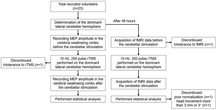
A total of 23 healthy volunteers were recruited, and 19 healthy volunteers were finally included for the statistical analysis. Before stimulation, the cerebellar hemisphere dominant for swallowing was determined by the single-pulse TMS. The cerebellar representation of the suprahyoid muscles of this hemisphere was selected as the target for stimulation with 10 Hz rTMS, 100% resting motor threshold (rMT), and 250 pulses, with every 1 s of stimulation followed by an interval of 9 s. The motor evoked potential (MEP) amplitude of the suprahyoid muscles in the bilateral cerebral cortex was measured before and after stimulation to evaluate the cortical excitability. Forty-eight hours after elution, rTMS was reapplied on the dominant cerebellar representation of the suprahyoid muscles with the same stimulation parameters. Rs-fMRI was performed before and after stimulation to observe the changes in amplitude of low-frequency fluctuation (ALFF) and regional homology (ReHo) at 0.01-0.08 Hz, 0.01-0.027 Hz, and 0.027-0.073 Hz.
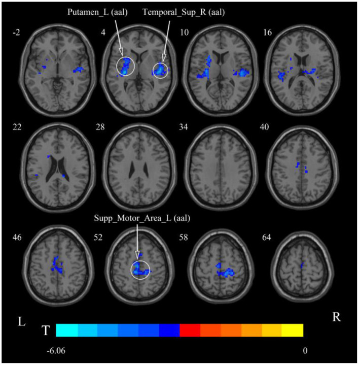
After cerebellar high-frequency rTMS, MEP recorded from swallowing-related bilateral cerebral cortex was increased. The results of rs-fMRI showed that at 0.01-0.08 Hz, ALFF was increased at the pons, right cerebellum, and medulla and decreased at the left temporal lobe, and ReHo was decreased at the left insular lobe, right temporal lobe, and corpus callosum. At 0.01-0.027 Hz, ALFF was decreased at the left temporal lobe, and ReHo was decreased at the right temporal lobe, left putamen, and left supplementary motor area.
Repetitive transcranial magnetic stimulation of the swallowing cortex in the dominant cerebellar hemisphere increased the bilateral cerebral swallowing cortex excitability and enhanced pontine, bulbar, and cerebellar spontaneous neural activity, suggesting that unilateral high-frequency stimulation of the cerebellum can excite both brainstem and cortical swallowing centers. These findings all provide favorable support for the application of cerebellar rTMS in the clinical practice.
采用静息态功能磁共振成像(rs-fMRI)研究小脑高频重复经颅磁刺激(rTMS)对吞咽相关神经网络的影响及可能机制。
共招募23名健康志愿者,最终纳入19名健康志愿者进行统计分析。刺激前,通过单脉冲TMS确定吞咽优势小脑半球。选择该半球舌骨上肌群的小脑代表区作为刺激靶点,采用10Hz rTMS、100%静息运动阈值(rMT)和250个脉冲进行刺激,每刺激1秒后间隔9秒。分别在刺激前后测量双侧大脑皮质舌骨上肌群的运动诱发电位(MEP)幅值,以评估皮质兴奋性。洗脱48小时后,采用相同刺激参数再次对舌骨上肌群优势小脑代表区进行rTMS刺激。在刺激前后进行rs-fMRI,观察0.01 - 0.08Hz、0.01 - 0.027Hz和0.027 - 0.073Hz频段低频振幅(ALFF)和局部一致性(ReHo)的变化。
小脑高频rTMS后,吞咽相关双侧大脑皮质记录的MEP幅值增加。rs-fMRI结果显示,在0.01 - 0.08Hz频段,脑桥、右侧小脑和延髓的ALFF增加,左侧颞叶的ALFF降低,左侧岛叶、右侧颞叶和胼胝体的ReHo降低。在0.01 - 0.027Hz频段,左侧颞叶的ALFF降低,右侧颞叶、左侧壳核和左侧辅助运动区的ReHo降低。
优势小脑半球吞咽皮质的重复经颅磁刺激增加了双侧大脑吞咽皮质的兴奋性,增强了脑桥、延髓和小脑的自发神经活动,提示单侧小脑高频刺激可兴奋脑干和皮质吞咽中枢。这些发现均为小脑rTMS在临床实践中的应用提供了有力支持。