Department of Electrical Engineering, National Taiwan University, Taipei, Taiwan, 106.
Department of Neurology, Taipei Veteran General Hospital, Taipei, Taiwan.
Neurotherapeutics. 2022 Jul;19(4):1368-1380. doi: 10.1007/s13311-022-01250-7. Epub 2022 May 17.
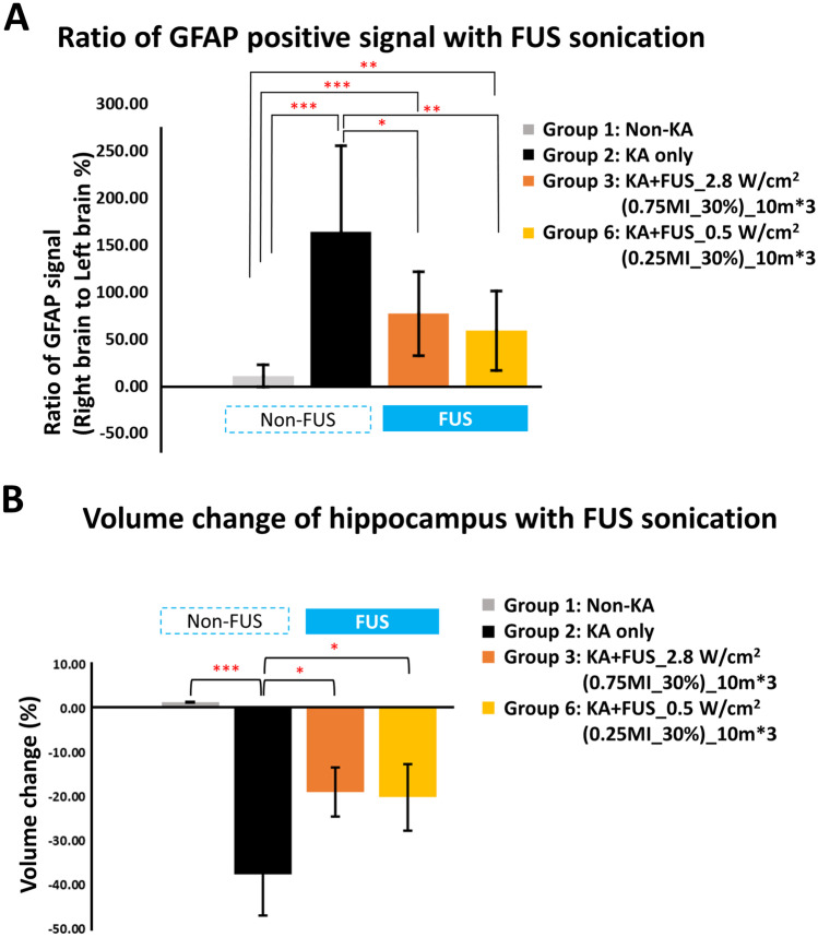
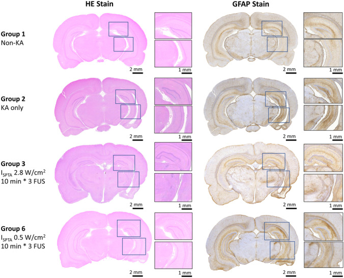
Focused ultrasound (FUS) has potential utility for modulating regional brain excitability and possibly aiding seizure control; however, the duration of any beneficial effect is unknown. This study explores the efficacy and time course of a short series of pulsed FUS in suppressing EEG epileptiform spikes/bursts in a kainic acid (KA) animal model of temporal lobe epilepsy. Forty-four male Sprague-Dawley rats were recorded for 14 weeks with EEG while software calculated EEG numbers of epileptiform spikes and bursts (≥ 3 spikes/s). Four regimens of FUS given in a single session at week 7 were evaluated, with mechanical index (MI) ranging from 0.25 to 0.75, intensity spatial peak temporal average (I) from 0.1 to 2.8 W per cm, duty cycle from 1 to 30%, and three consecutive pulse trains for 5 or 10 min each. Controls included sham injections in four and KA without FUS in eleven animals. Histological analysis investigated tissue effects. All animals receiving KA evidenced EEG spikes, averaging 10,378 ± 1651 spikes per 8 h and 1255 ± 199 bursts per 8 h by weeks 6-7. The KA-only group showed a 30% of increase in spikes and bursts by week 14. Compared to the KA-only group, spike counts were reduced by about 25%, burst counts by about 33%, and burst durations by about 50% with FUS. Behavioral seizures were not analyzed, but electrographic seizures longer than 10 s declined up to 70% after some FUS regimens. Repeated-measure ANOVA showed a significant effect of higher intensity and longer sonication duration FUS treatment using 0.75-MI, I 2.8 W/cm, 30% duty cycle for 10-min sonications (group effect, F (4, 15) = 6.321, p < 0.01; interaction effect, F (44, 165) = 1.726, p < 0.01), with the hippocampal protective effect lasting to week 14, accompanied by decreased inflammation and gliosis effect. In contrast, spike and burst suppression were achieved using an FUS regimen with 0.25-MI I 0.5 W/cm, 30% duty cycle for 10-min sonications. This regimen reduced inflammation and gliosis at weeks 8-14 and protected hippocampal tissue. This study demonstrates that low-intensity pulsed ultrasound can modulate epileptiform activity for up to 7 weeks and, if replicated in the clinical setting, might be a practical treatment for epilepsy.
聚焦超声(FUS)在调节局部脑兴奋性和可能辅助控制癫痫发作方面具有潜在的应用价值;然而,其任何有益效果的持续时间尚不清楚。本研究探讨了一系列短时间脉冲 FUS 在抑制颞叶癫痫动物模型中海马区癫痫样电爆发/爆发中的疗效和时间过程。44 只雄性 Sprague-Dawley 大鼠在接受脑电图记录的 14 周内,软件计算出癫痫样电爆发/爆发的脑电图数量(≥3 个/秒)。评估了在第 7 周单次治疗中进行的四种 FUS 方案,机械指数(MI)范围为 0.25 至 0.75,强度空间峰值时间平均(I)为 0.1 至 2.8 W/cm,占空比为 1 至 30%,连续三个脉冲串,每个脉冲串持续 5 或 10 分钟。对照组包括 4 只接受假注射的动物和 11 只未接受 FUS 的 KA 动物。组织学分析研究了组织效应。所有接受 KA 的动物在第 6-7 周均出现脑电图棘波,平均每 8 小时出现 10378±1651 个棘波和 1255±199 个爆发。仅 KA 组在第 14 周时棘波和爆发增加了 30%。与仅 KA 组相比,FUS 可使棘波计数减少约 25%,爆发计数减少约 33%,爆发持续时间减少约 50%。未分析行为性癫痫发作,但一些 FUS 方案后超过 10 秒的电发作减少了高达 70%。重复测量方差分析显示,使用 0.75-MI、I 2.8 W/cm、30%占空比 10 分钟超声治疗的高强度和更长超声持续时间的 FUS 治疗具有显著效果(组间效应,F(4,15)=6.321,p<0.01;交互效应,F(44,165)=1.726,p<0.01),其海马保护作用持续到第 14 周,伴有炎症和神经胶质增生减少。相比之下,使用 0.25-MI I 0.5 W/cm、30%占空比 10 分钟超声的 FUS 方案可实现棘波和爆发的抑制。该方案在第 8-14 周减少了炎症和神经胶质增生,并保护了海马组织。本研究表明,低强度脉冲超声可调节癫痫样活动长达 7 周,如果在临床环境中得到复制,可能成为一种实用的癫痫治疗方法。