Department of Molecular Biotechnology and Health Sciences, University of Turin, Turin, Italy.
Department of Systems Biology and Bioinformatics, University of Rostock, Rostock, Germany.
J Extracell Vesicles. 2022 May;11(5):e12217. doi: 10.1002/jev2.12217.
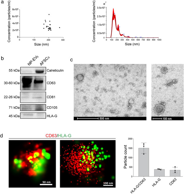
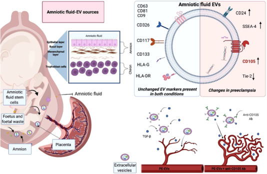
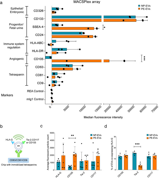
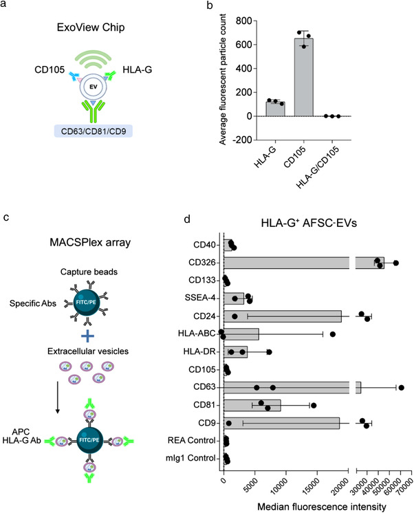
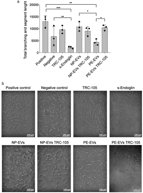
Amniotic fluid surrounding the developing fetus is a complex biological fluid rich in metabolically active bio-factors. The presence of extracellular vesicles (EVs) in amniotic fluid has been mainly related to foetal urine. We here characterized EVs from term amniotic fluid in terms of surface marker expression using different orthogonal techniques. EVs appeared to be a heterogeneous population expressing markers of renal, placental, epithelial and stem cells. Moreover, we compared amniotic fluid EVs from normal pregnancies with those of preeclampsia, a hypertensive disorder affecting up to 8% of pregnancies worldwide. An increase of CD105 (endoglin) expressing EVs was observed in preeclamptic amniotic fluid by bead-based cytofluorimetric analysis, and further confirmed using a chip-based analysis. HLA-G, a typical placental marker, was not co-expressed by the majority of CD105 EVs, in analogy with amniotic fluid stromal cell derived-EVs. At a functional level, preeclampsia-derived EVs, but not normal pregnancy EVs, showed an antiangiogenic effect, possibly due to the decoy effect of endoglin. Our results provide a characterization of term amniotic fluid-EVs, supporting their origin from foetal and placental cells. In preeclampsia, the observed antiangiogenic characteristics of amniotic fluid-EVs may reflect the hypoxic and antiangiogenic microenvironment and could possibly impact on the developing fetus or on the surrounding foetal membranes.
羊水是环绕在发育中胎儿周围的一种复杂的生物液体,富含代谢活跃的生物因子。羊水中外泌体(EVs)的存在主要与胎儿尿液有关。我们使用不同的正交技术,从足月羊水的 EVs 方面对其表面标记物表达进行了特征描述。EVs 似乎是一个异质群体,表达肾、胎盘、上皮和干细胞的标记物。此外,我们比较了正常妊娠和先兆子痫(一种影响全球多达 8%妊娠的高血压疾病)患者的羊水 EVs。通过基于珠子的细胞荧光分析观察到先兆子痫羊水的 CD105(内皮糖蛋白)表达 EVs 增加,并使用基于芯片的分析进一步证实。HLA-G,一种典型的胎盘标记物,没有被大多数 CD105 EVs 共同表达,与羊水基质细胞衍生的 EVs 类似。在功能水平上,子痫前期衍生的 EVs,但不是正常妊娠的 EVs,显示出抗血管生成作用,可能是由于内皮糖蛋白的诱饵作用。我们的结果提供了足月羊水-EVs 的特征描述,支持它们来源于胎儿和胎盘细胞。在先兆子痫中,羊水-EVs 的观察到的抗血管生成特征可能反映了缺氧和抗血管生成的微环境,并可能对发育中的胎儿或周围的胎膜产生影响。