Retinal and Inflammatory Eye Diseases, Centre for Ophthalmic Specialized Care (COS), Clinic Montchoisi Teaching Centre, Lausanne, Switzerland.
Department of Ophthalmology, Istanbul Faculty of Medicine, Istanbul University, Istanbul, Turkey.
Eye (Lond). 2021 Jan;35(1):52-73. doi: 10.1038/s41433-020-1072-0. Epub 2020 Aug 10.
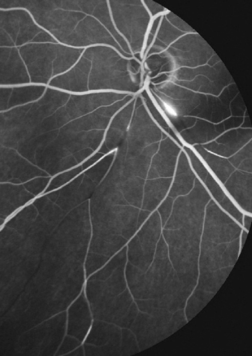
The aim of this review was to identify the imaging methods at our disposal to optimally manage posterior uveitis at the present time. The focus was put on methods that have become available since the 1990s, some 30 years after fluorescein angiography had revolutionised imaging of posterior uveitis in particular imaging of the retinal vascular structures in the 1960s. We have focussed our review on precise imaging methods that have been standardised and validated and can be used universally thanks to commercially produced and available instruments for the diagnosis and follow-up of posterior uveitis. The second part of this imaging review will deal with invasive imaging methods and in particular ocular angiography.
本次综述的目的是确定目前可用于优化后葡萄膜炎管理的影像学方法。重点放在自 20 世纪 90 年代以来出现的方法上,即荧光素血管造影术在 20 世纪 60 年代特别革新了后葡萄膜炎的影像学,包括视网膜血管结构的影像学之后大约 30 年。我们的综述集中在已经标准化和验证的精确成像方法上,由于商业生产和可用于后葡萄膜炎诊断和随访的仪器,这些方法可以在全球范围内使用。本影像学综述的第二部分将涉及有创影像学方法,特别是眼血管造影术。