文献检索文档翻译深度研究
Suppr Zotero 插件Zotero 插件
邀请有礼套餐&价格历史记录

新学期,新优惠

限时优惠:9月1日-9月22日

30天高级会员仅需29元

1天体验卡首发特惠仅需5.99元

了解详情
不再提醒
插件&应用
Suppr Zotero 插件Zotero 插件浏览器插件Mac 客户端Windows 客户端微信小程序
高级版
套餐订阅购买积分包
AI 工具
文献检索文档翻译深度研究
关于我们
关于 Suppr公司介绍联系我们用户协议隐私条款
关注我们

Suppr 超能文献

核心技术专利:CN118964589B侵权必究
粤ICP备2023148730 号-1Suppr @ 2025

SARS-CoV-2 宿主关闭会影响先天 NK 细胞的功能,但通过非刺突抗体强烈激活抗体依赖性 NK 活性。

SARS-CoV-2 host-shutoff impacts innate NK cell functions, but antibody-dependent NK activity is strongly activated through non-spike antibodies.

机构信息

Division of Infection and Immunity, School of Medicine, Cardiff University, Cardiff, United Kingdom.

Cambridge Institute for Therapeutic Immunology & Infectious Disease, Jeffrey Cheah Biomedical Centre, Cambridge Biomedical Campus, University of Cambridge, Cambridge, United Kingdom.

出版信息

Elife. 2022 May 19;11:e74489. doi: 10.7554/eLife.74489.


DOI:10.7554/eLife.74489
PMID:35587364
原文链接:https://pmc.ncbi.nlm.nih.gov/articles/PMC9239683/
Abstract

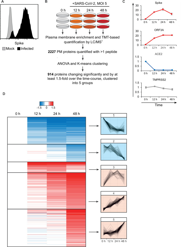
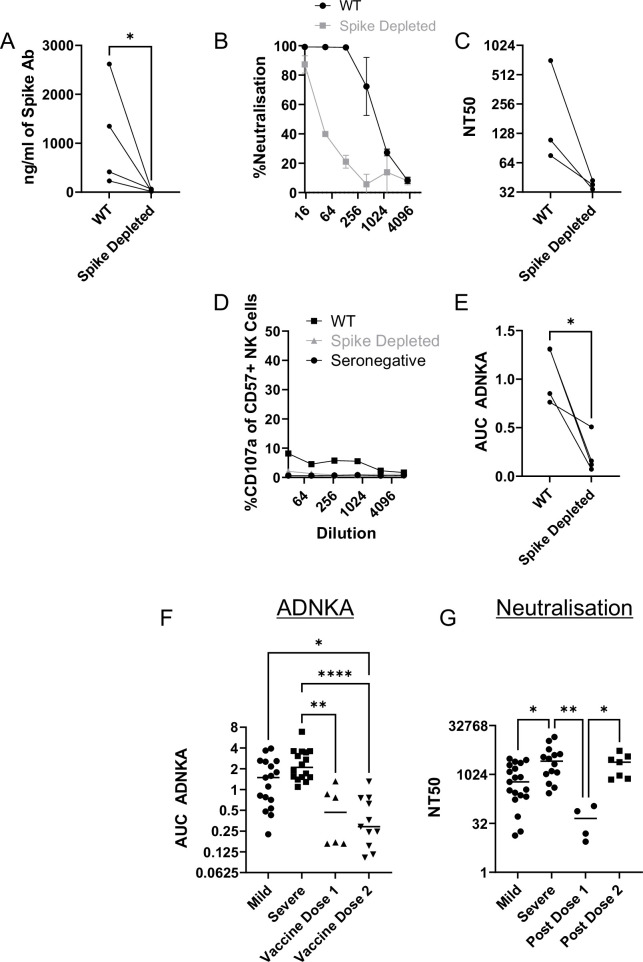
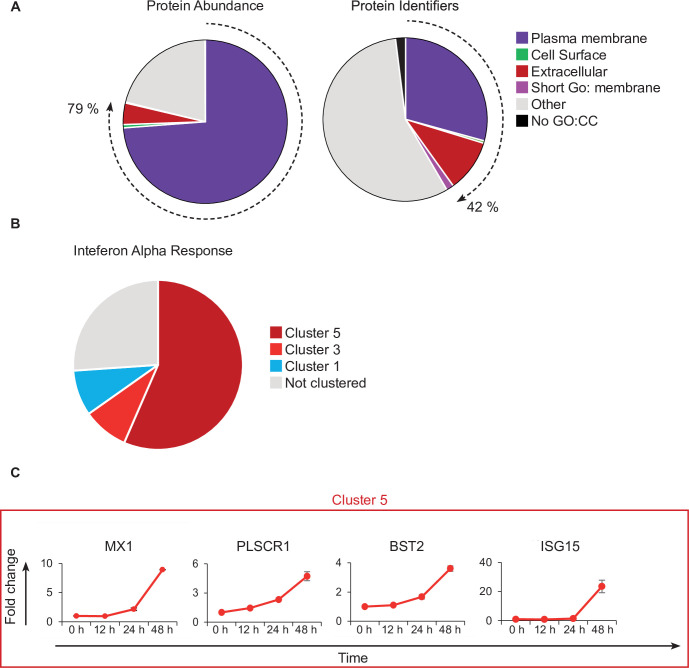
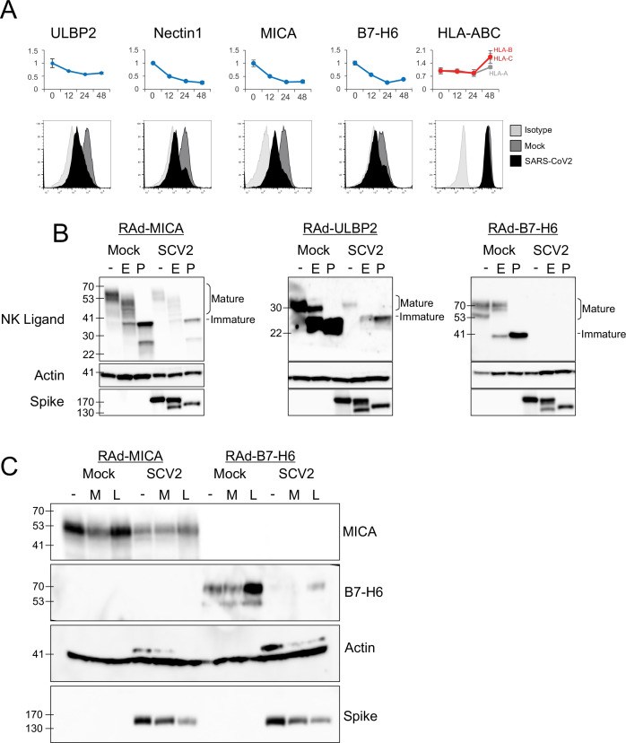
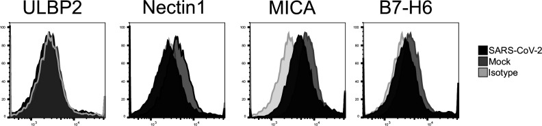
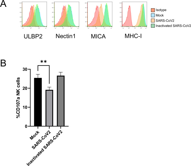
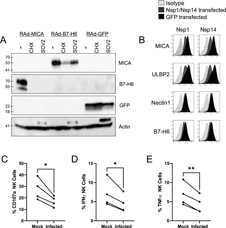
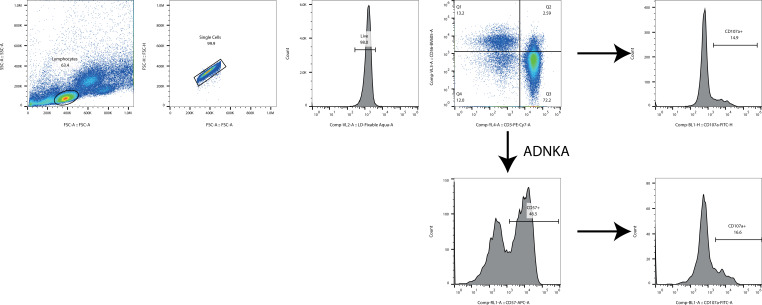
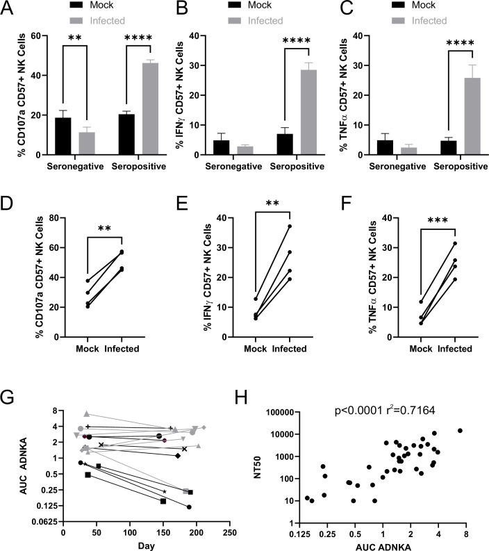
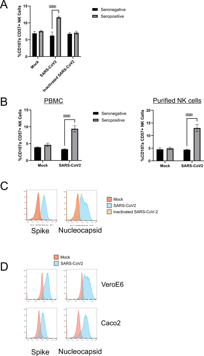
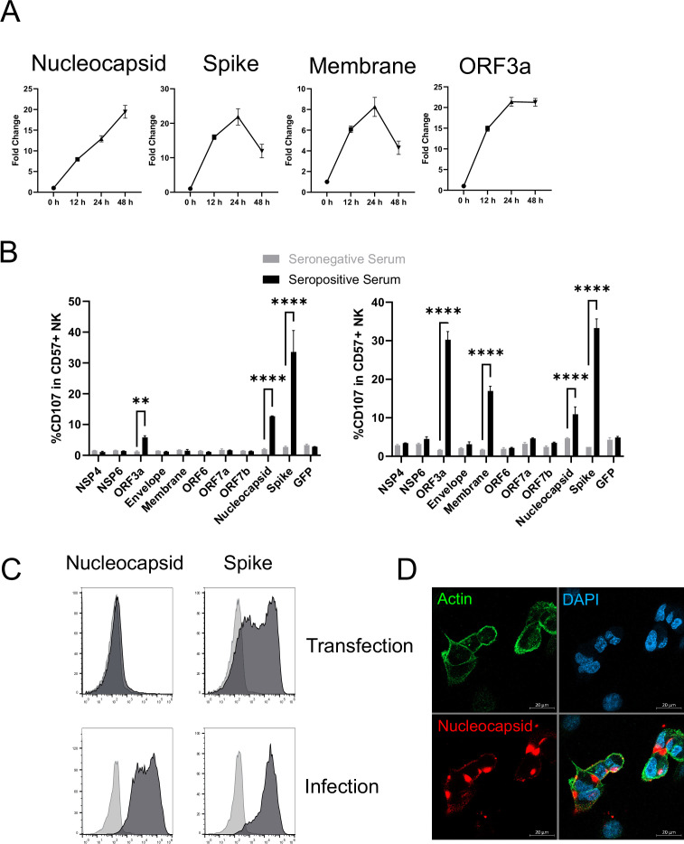
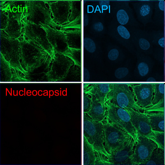
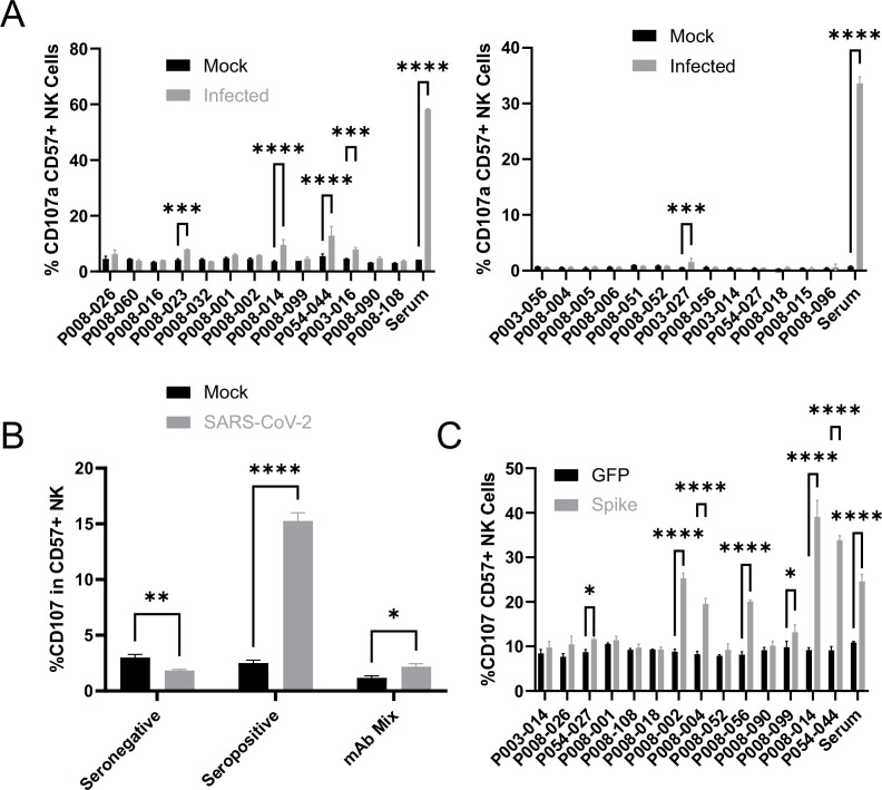
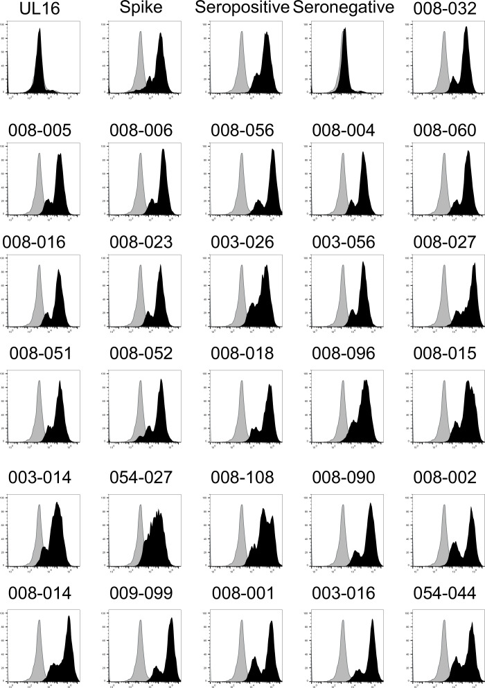
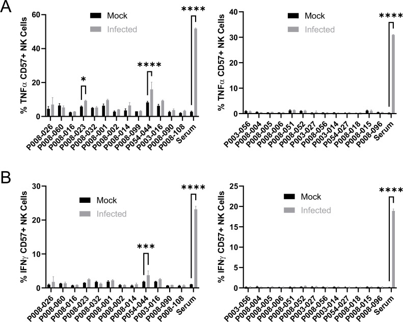
The outcome of infection is dependent on the ability of viruses to manipulate the infected cell to evade immunity, and the ability of the immune response to overcome this evasion. Understanding this process is key to understanding pathogenesis, genetic risk factors, and both natural and vaccine-induced immunity. SARS-CoV-2 antagonises the innate interferon response, but whether it manipulates innate cellular immunity is unclear. An unbiased proteomic analysis determined how cell surface protein expression is altered on SARS-CoV-2-infected lung epithelial cells, showing downregulation of activating NK ligands B7-H6, MICA, ULBP2, and Nectin1, with minimal effects on MHC-I. This occurred at the level of protein synthesis, could be mediated by Nsp1 and Nsp14, and correlated with a reduction in NK cell activation. This identifies a novel mechanism by which SARS-CoV-2 host-shutoff antagonises innate immunity. Later in the disease process, strong antibody-dependent NK cell activation (ADNKA) developed. These responses were sustained for at least 6 months in most patients, and led to high levels of pro-inflammatory cytokine production. Depletion of spike-specific antibodies confirmed their dominant role in neutralisation, but these antibodies played only a minor role in ADNKA compared to antibodies to other proteins, including ORF3a, Membrane, and Nucleocapsid. In contrast, ADNKA induced following vaccination was focussed solely on spike, was weaker than ADNKA following natural infection, and was not boosted by the second dose. These insights have important implications for understanding disease progression, vaccine efficacy, and vaccine design.

摘要

病毒操纵感染细胞逃避免疫以及免疫反应克服这种逃避的能力决定了感染的结果。了解这一过程是理解发病机制、遗传风险因素以及自然和疫苗诱导免疫的关键。SARS-CoV-2 拮抗先天干扰素反应,但它是否操纵先天细胞免疫尚不清楚。一项无偏蛋白质组学分析确定了 SARS-CoV-2 感染的肺上皮细胞表面蛋白表达如何改变,显示出激活 NK 配体 B7-H6、MICA、ULBP2 和 Nectin1 的下调,而 MHC-I 的影响最小。这发生在蛋白质合成水平,可以由 Nsp1 和 Nsp14 介导,并与 NK 细胞激活减少相关。这确定了 SARS-CoV-2 宿主关闭拮抗先天免疫的新机制。在疾病过程的后期,出现了强烈的抗体依赖性 NK 细胞激活(ADNKA)。这些反应在大多数患者中至少持续了 6 个月,并导致高水平的促炎细胞因子产生。耗尽刺突特异性抗体证实了它们在中和中的主导作用,但与其他蛋白(包括 ORF3a、Membrane 和 Nucleocapsid)的抗体相比,这些抗体在 ADNKA 中的作用较小。相比之下,接种疫苗后诱导的 ADNKA 仅针对刺突,比自然感染后诱导的 ADNKA 弱,第二剂不会增强。这些见解对理解疾病进展、疫苗效力和疫苗设计具有重要意义。

https://cdn.ncbi.nlm.nih.gov/pmc/blobs/d128/9239683/fd70cd854f76/elife-74489-fig9.jpg
https://cdn.ncbi.nlm.nih.gov/pmc/blobs/d128/9239683/df19a7d8c17c/elife-74489-fig1.jpg
https://cdn.ncbi.nlm.nih.gov/pmc/blobs/d128/9239683/a9fb9ae67a19/elife-74489-fig1-figsupp1.jpg
https://cdn.ncbi.nlm.nih.gov/pmc/blobs/d128/9239683/bde6e686b73c/elife-74489-fig1-figsupp2.jpg
https://cdn.ncbi.nlm.nih.gov/pmc/blobs/d128/9239683/46f61b274645/elife-74489-fig1-figsupp3.jpg
https://cdn.ncbi.nlm.nih.gov/pmc/blobs/d128/9239683/5d26b80fdf07/elife-74489-fig2.jpg
https://cdn.ncbi.nlm.nih.gov/pmc/blobs/d128/9239683/ba2f43e4129f/elife-74489-fig2-figsupp1.jpg
https://cdn.ncbi.nlm.nih.gov/pmc/blobs/d128/9239683/73744585062c/elife-74489-fig3.jpg
https://cdn.ncbi.nlm.nih.gov/pmc/blobs/d128/9239683/29b6130a5bb9/elife-74489-fig3-figsupp1.jpg
https://cdn.ncbi.nlm.nih.gov/pmc/blobs/d128/9239683/76864fa76b1a/elife-74489-fig3-figsupp2.jpg
https://cdn.ncbi.nlm.nih.gov/pmc/blobs/d128/9239683/0a26f66ae8e1/elife-74489-fig4.jpg
https://cdn.ncbi.nlm.nih.gov/pmc/blobs/d128/9239683/b0f569e278a9/elife-74489-fig4-figsupp1.jpg
https://cdn.ncbi.nlm.nih.gov/pmc/blobs/d128/9239683/0558ea322675/elife-74489-fig5.jpg
https://cdn.ncbi.nlm.nih.gov/pmc/blobs/d128/9239683/0cfea7dfdb96/elife-74489-fig5-figsupp1.jpg
https://cdn.ncbi.nlm.nih.gov/pmc/blobs/d128/9239683/7c5b6fce07da/elife-74489-fig6.jpg
https://cdn.ncbi.nlm.nih.gov/pmc/blobs/d128/9239683/966ebb76bab7/elife-74489-fig6-figsupp1.jpg
https://cdn.ncbi.nlm.nih.gov/pmc/blobs/d128/9239683/b6e0b34ff8bd/elife-74489-fig7.jpg
https://cdn.ncbi.nlm.nih.gov/pmc/blobs/d128/9239683/16c8e593e675/elife-74489-fig7-figsupp1.jpg
https://cdn.ncbi.nlm.nih.gov/pmc/blobs/d128/9239683/9c478ab6ab61/elife-74489-fig7-figsupp2.jpg
https://cdn.ncbi.nlm.nih.gov/pmc/blobs/d128/9239683/4cbf4e7838ec/elife-74489-fig8.jpg
https://cdn.ncbi.nlm.nih.gov/pmc/blobs/d128/9239683/fd70cd854f76/elife-74489-fig9.jpg

相似文献

[1]
SARS-CoV-2 host-shutoff impacts innate NK cell functions, but antibody-dependent NK activity is strongly activated through non-spike antibodies.

Elife. 2022-5-19

[2]
Natural killer cell-mediated ADCC in SARS-CoV-2-infected individuals and vaccine recipients.

Eur J Immunol. 2022-8

[3]
Siglec-9 Restrains Antibody-Dependent Natural Killer Cell Cytotoxicity against SARS-CoV-2.

mBio. 2023-2-28

[4]
Impact of SARS-CoV-2 vaccination on FcγRIIIA/CD16 dynamics in Natural Killer cells: relevance for antibody-dependent functions.

Front Immunol. 2023

[5]
Antibody-dependent NK-cell and neutralizing antibody responses against the Spike protein of Wuhan-Hu-1 and Omicron BA.1 SARS-CoV-2 variants in vaccinated experienced and vaccinated naïve individuals.

J Med Virol. 2023-7

[6]
SARS-CoV-2 escapes direct NK cell killing through Nsp1-mediated downregulation of ligands for NKG2D.

Cell Rep. 2022-12-27

[7]
Innate Immune Responses of Vaccinees Determine Early Neutralizing Antibody Production After ChAdOx1nCoV-19 Vaccination.

Front Immunol. 2022

[8]
Emerging Variants of SARS-CoV-2 and Novel Therapeutics Against Coronavirus (COVID-19)

2025-1

[9]
Natural killer cell responses during SARS-CoV-2 infection and vaccination in people living with HIV-1.

Sci Rep. 2023-11-3

[10]
mRNA vaccine-induced T cells respond identically to SARS-CoV-2 variants of concern but differ in longevity and homing properties depending on prior infection status.

Elife. 2021-10-12

引用本文的文献

[1]
An Autoimmunity-associated allele of enhances the innate immune response to promote protection against acute coronavirus infection.

bioRxiv. 2025-7-18

[2]
Protective Potential and Functional Role of Antibodies Against SARS-CoV-2 Nucleocapsid Protein.

Antibodies (Basel). 2025-5-28

[3]
Identification of soluble biomarkers that associate with distinct manifestations of long COVID.

Nat Immunol. 2025-5

[4]
Cell surface RNA virus nucleocapsid proteins: a viral strategy for immunosuppression?

Npj Viruses. 2024-9-2

[5]
Chemokines simultaneously bind SARS-CoV-2 nucleocapsid protein RNA-binding and dimerization domains.

Virol J. 2025-3-17

[6]
From natural defenders to therapeutic warriors: NK cells in HIV immunotherapy.

Immunotherapy. 2025-2

[7]
Detrimental Effects of Anti-Nucleocapsid Antibodies in SARS-CoV-2 Infection, Reinfection, and the Post-Acute Sequelae of COVID-19.

Pathogens. 2024-12-15

[8]
Longitudinal transcriptional changes reveal genes from the natural killer cell-mediated cytotoxicity pathway as critical players underlying COVID-19 progression.

Elife. 2024-10-29

[9]
Delineating the functional activity of antibodies with cross-reactivity to SARS-CoV-2, SARS-CoV-1 and related sarbecoviruses.

PLoS Pathog. 2024-10

[10]
Sequence Matters: Primary COVID-19 Vaccination after Infection Elicits Similar Anti-spike Antibody Levels, but Stronger Antibody Dependent Cell-mediated Cytotoxicity than Breakthrough Infection.

J Immunol. 2024-10-15

本文引用的文献

[1]
Functional convalescent plasma antibodies and pre-infusion titers shape the early severe COVID-19 immune response.

Nat Commun. 2021-11-25

[2]
Untimely TGFβ responses in COVID-19 limit antiviral functions of NK cells.

Nature. 2021-12

[3]
Coronavirus Nsp1: Immune Response Suppression and Protein Expression Inhibition.

Front Microbiol. 2021-9-28

[4]
Fc-engineered antibody therapeutics with improved anti-SARS-CoV-2 efficacy.

Nature. 2021-11

[5]
Fab and Fc contribute to maximal protection against SARS-CoV-2 following NVX-CoV2373 subunit vaccine with Matrix-M vaccination.

Cell Rep Med. 2021-9-21

[6]
The Fc-mediated effector functions of a potent SARS-CoV-2 neutralizing antibody, SC31, isolated from an early convalescent COVID-19 patient, are essential for the optimal therapeutic efficacy of the antibody.

PLoS One. 2021

[7]
The landscape of antibody binding in SARS-CoV-2 infection.

PLoS Biol. 2021-6

[8]
A single dose of the SARS-CoV-2 vaccine BNT162b2 elicits Fc-mediated antibody effector functions and T cell responses.

Cell Host Microbe. 2021-7-14

[9]
Contribution of SARS-CoV-2 Accessory Proteins to Viral Pathogenicity in K18 Human ACE2 Transgenic Mice.

J Virol. 2021-8-10

[10]
Translational shutdown and evasion of the innate immune response by SARS-CoV-2 NSP14 protein.

Proc Natl Acad Sci U S A. 2021-6-15

文献AI研究员

20分钟写一篇综述,助力文献阅读效率提升50倍

立即体验

用中文搜PubMed

大模型驱动的PubMed中文搜索引擎

马上搜索

推荐工具

医学文档翻译智能文献检索