Proteomics Program, Novo Nordisk Foundation Center for Protein Research, Faculty of Health and Medical Sciences, University of Copenhagen, Copenhagen, Denmark.
Spatial Proteomics Group, Max Delbrück Center for Molecular Medicine in the Helmholtz Association, Berlin, Germany.
Nat Biotechnol. 2022 Aug;40(8):1231-1240. doi: 10.1038/s41587-022-01302-5. Epub 2022 May 19.
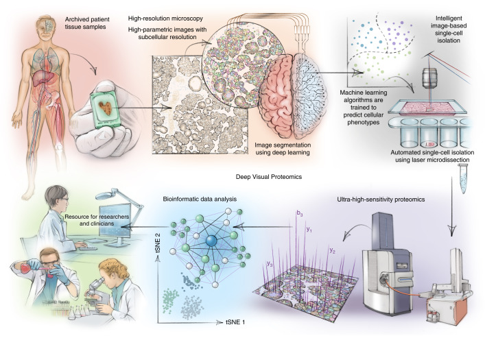
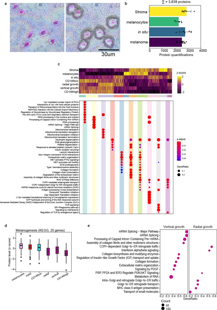
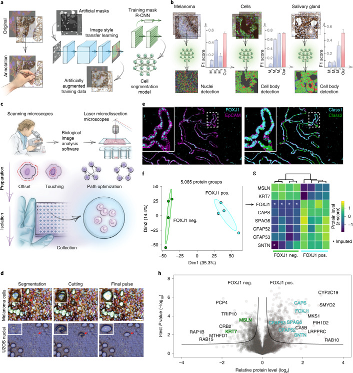
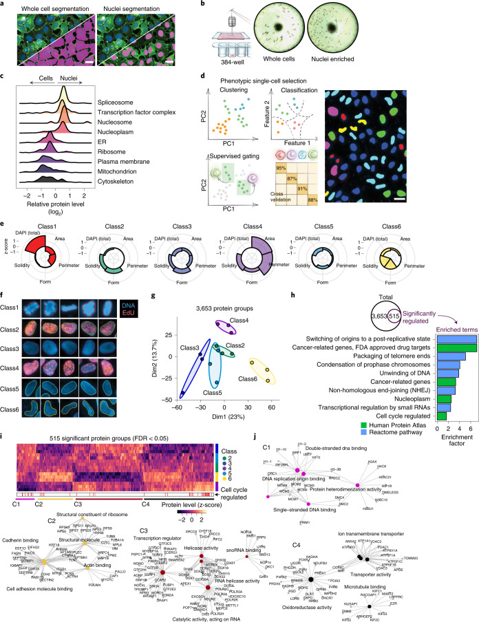
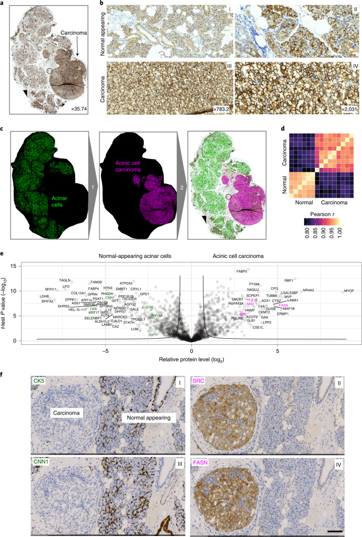
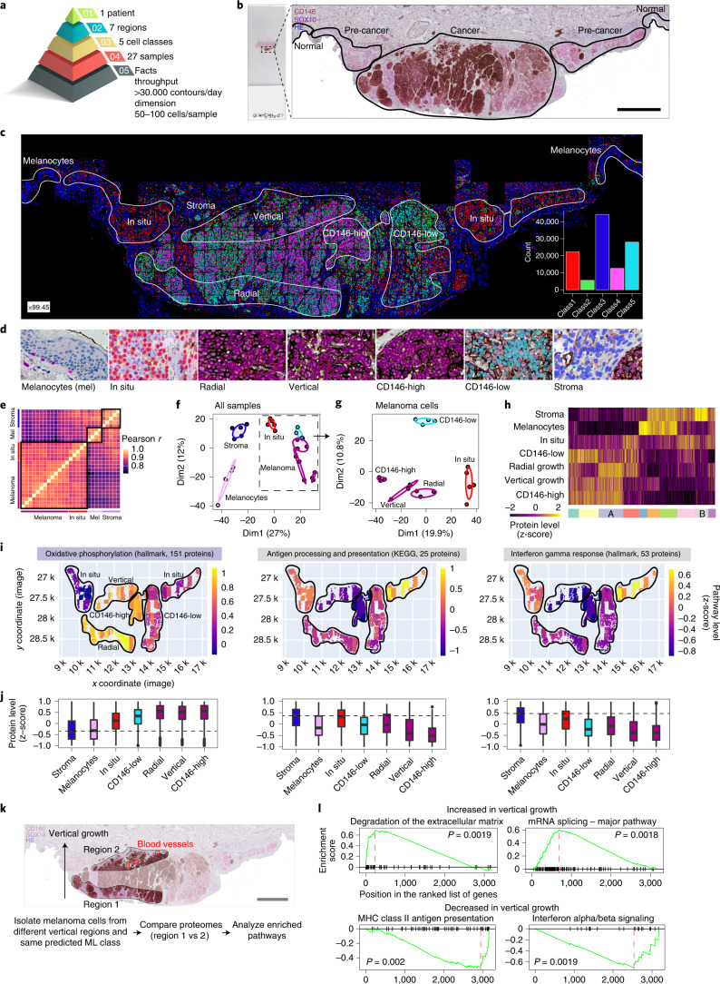
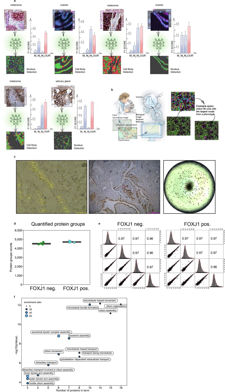
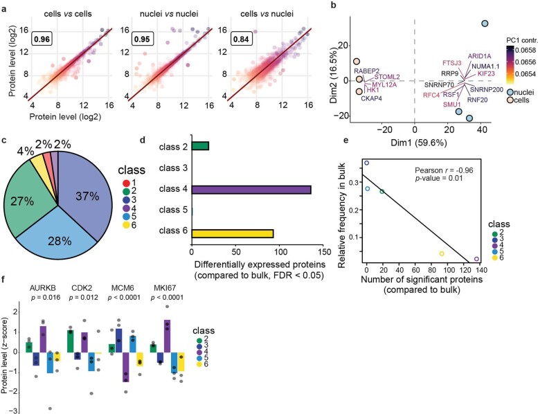
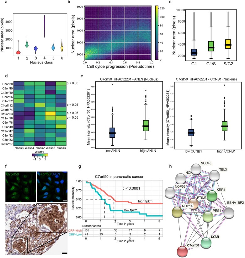
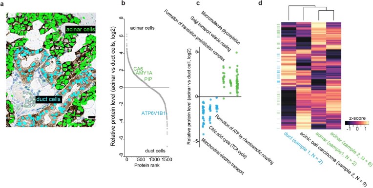
Despite the availabilty of imaging-based and mass-spectrometry-based methods for spatial proteomics, a key challenge remains connecting images with single-cell-resolution protein abundance measurements. Here, we introduce Deep Visual Proteomics (DVP), which combines artificial-intelligence-driven image analysis of cellular phenotypes with automated single-cell or single-nucleus laser microdissection and ultra-high-sensitivity mass spectrometry. DVP links protein abundance to complex cellular or subcellular phenotypes while preserving spatial context. By individually excising nuclei from cell culture, we classified distinct cell states with proteomic profiles defined by known and uncharacterized proteins. In an archived primary melanoma tissue, DVP identified spatially resolved proteome changes as normal melanocytes transition to fully invasive melanoma, revealing pathways that change in a spatial manner as cancer progresses, such as mRNA splicing dysregulation in metastatic vertical growth that coincides with reduced interferon signaling and antigen presentation. The ability of DVP to retain precise spatial proteomic information in the tissue context has implications for the molecular profiling of clinical samples.
尽管存在基于成像和质谱的空间蛋白质组学方法,但仍存在一个关键挑战,即将图像与单细胞分辨率的蛋白质丰度测量联系起来。在这里,我们介绍了深度视觉蛋白质组学(DVP),它将基于人工智能的细胞表型图像分析与自动化的单细胞或单个细胞核激光显微切割和超高灵敏度质谱结合在一起。DVP 将蛋白质丰度与复杂的细胞或亚细胞表型联系起来,同时保留空间背景。通过从细胞培养物中单独切除核,我们使用已知和未表征的蛋白质定义的蛋白质组谱对不同的细胞状态进行了分类。在一个存档的原发性黑色素瘤组织中,DVP 鉴定了空间分辨率的蛋白质组变化,因为正常黑素细胞向完全侵袭性黑色素瘤转变,揭示了随着癌症进展而以空间方式改变的途径,例如在垂直生长的转移性中 mRNA 剪接失调,这与干扰素信号和抗原呈递减少同时发生。DVP 在组织背景中保留精确空间蛋白质组信息的能力对临床样本的分子分析具有重要意义。