Fleming Martinez Alicia K, Döppler Heike R, Bastea Ligia I, Edenfield Brandy H, Liou Geou-Yarh, Storz Peter
Department of Cancer Biology, Mayo Clinic, 4500 San Pablo Road, Jacksonville, FL 32224, USA.
Department of Biological Sciences, Center for Cancer Research & Therapeutic Development, Clark Atlanta University, Atlanta, GA 30314, USA.
iScience. 2022 Apr 29;25(5):104327. doi: 10.1016/j.isci.2022.104327. eCollection 2022 May 20.
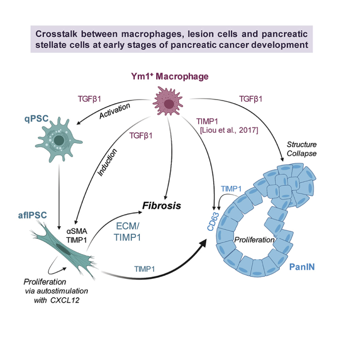
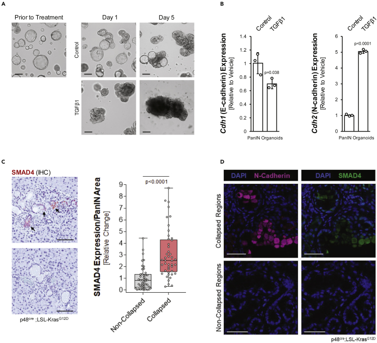
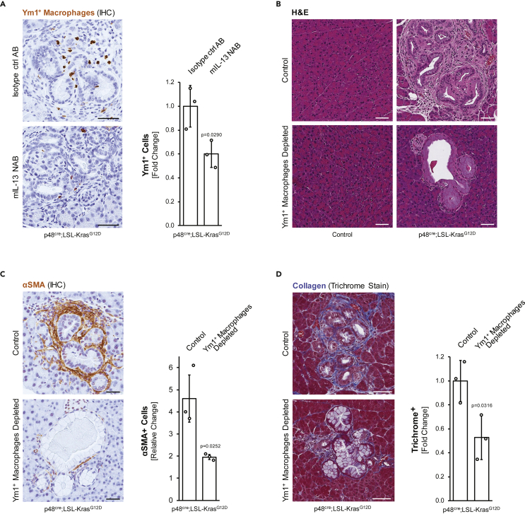
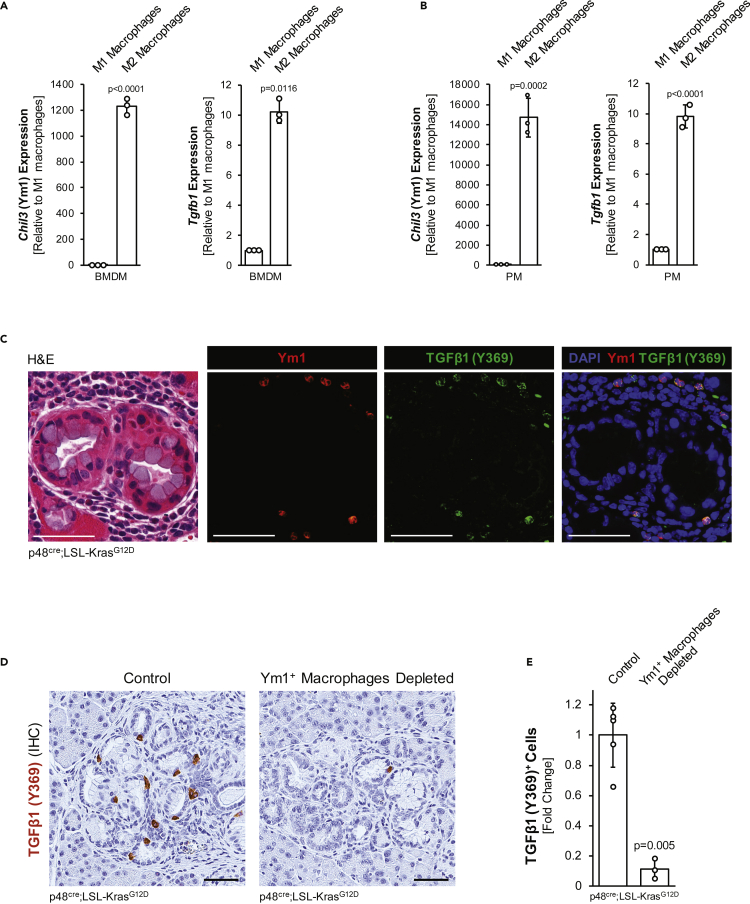
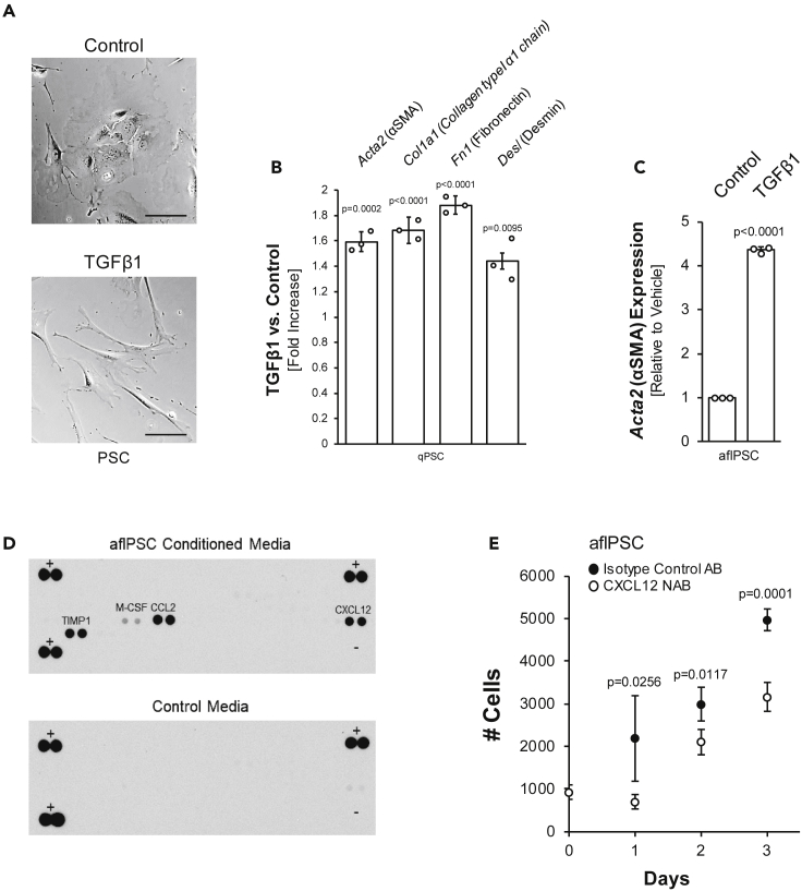
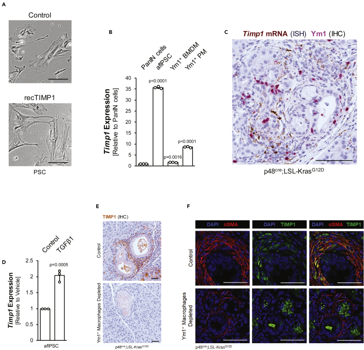
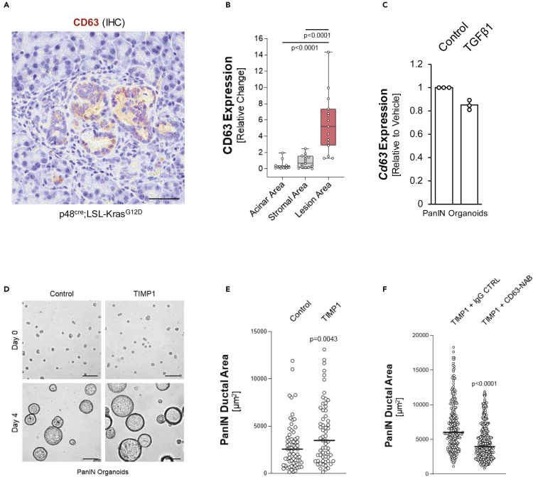
Desmoplasia around pancreatic lesions is a barrier for immune cells and a hallmark of developing and established pancreatic cancer. However, the contribution of the innate immune system to this process is ill-defined. Using the KC mouse model and primary cells , we show that alternatively activated macrophages (AAM) crosstalk with pancreatic lesion cells and pancreatic stellate cells (PSCs) to mediate fibrosis and progression of lesions. TGFβ1 secreted by AAM not only drives activation of quiescent PSCs but also in activated PSCs upregulates expression of TIMP1, a factor previously shown as crucial in fibrosis. Once activated, PSCs auto-stimulate proliferation via CXCL12. Furthermore, we found that TIMP1/CD63 signaling mediates PanIN lesion growth and TGFβ1 contributes to a cadherin switch and drives structural collapse of lesions, indicating a potential progression step. Taken together, our data indicate TGFβ1 produced by Ym1+ AAM as a major driver of processes that initiate the development of pancreatic cancer.
胰腺病变周围的促纤维增生是免疫细胞的一道屏障,也是胰腺癌发生发展过程中的一个标志。然而,固有免疫系统在这一过程中的作用尚不明确。利用KC小鼠模型和原代细胞,我们发现交替活化的巨噬细胞(AAM)与胰腺病变细胞及胰腺星状细胞(PSC)相互作用,介导纤维化和病变进展。AAM分泌的转化生长因子β1(TGFβ1)不仅驱动静止PSC的活化,而且在活化的PSC中上调金属蛋白酶组织抑制因子1(TIMP1)的表达,TIMP1是先前已证明在纤维化过程中起关键作用的一个因子。一旦被激活,PSC通过CXC趋化因子配体12(CXCL12)自我刺激增殖。此外,我们发现TIMP1/CD63信号传导介导胰腺上皮内瘤变(PanIN)病变生长,TGFβ1促成钙黏蛋白转换并驱动病变的结构塌陷,表明这是一个潜在的进展步骤。综上所述,我们的数据表明Ym1+ AAM产生的TGFβ1是启动胰腺癌发生过程的主要驱动因素。