Institute of Radiation Medicine, Shanghai Medical College, Fudan University, Shanghai, 200032, China.
D1 Medical Technology Company, Shanghai, 201802, China.
Stem Cell Res Ther. 2022 May 26;13(1):219. doi: 10.1186/s13287-022-02902-3.
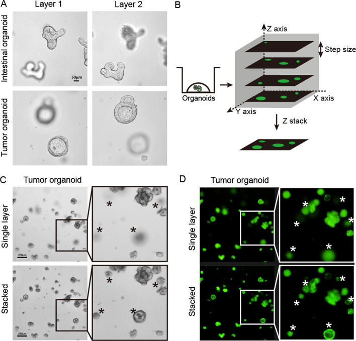
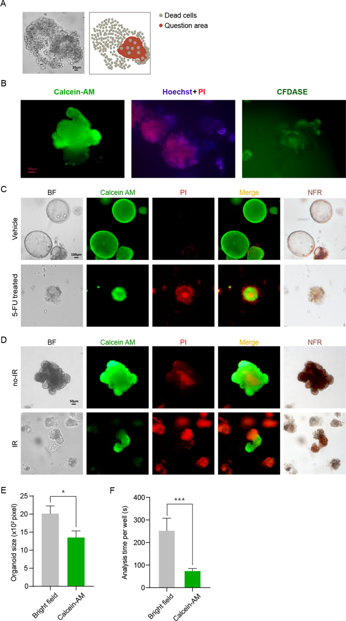
Organoids are three-dimensional structures that closely recapitulate tissue architecture and cellular composition, thereby holding great promise for organoid-based drug screening. Although growing in three-dimensional provides the possibility for organoids to recapitulate main features of corresponding tissues, it makes it incommodious for imaging organoids in two-dimensional and identifying surviving organoids from surrounding dead cells after organoids being treated by irradiation or chemotherapy. Therefore, significant work remains to establish high-quality controls to standardize organoid analyses and make organoid models more reproducible.
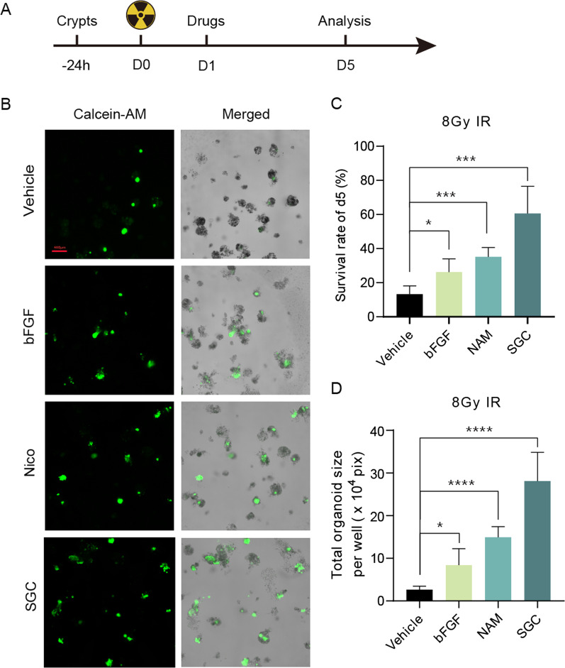
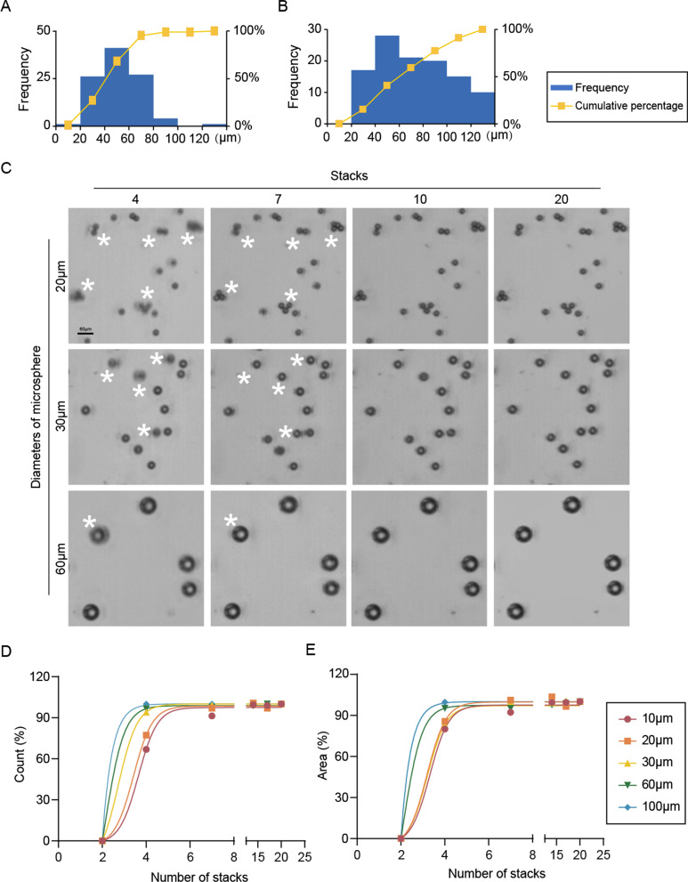
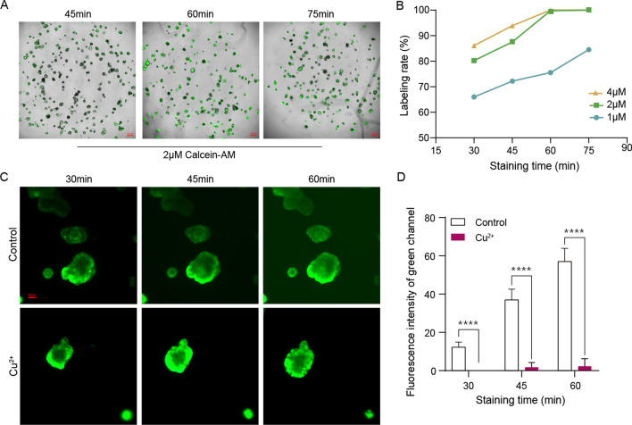
In this study, the Z-stack imaging technique was used for the imaging of three-dimensional organoids to gather all the organoids' maximum cross sections in one imaging. The combination of live cell staining fluorescent dye Calcein-AM and ImageJ assessment was used to analyze the survival of organoids treated by irradiation or chemotherapy.
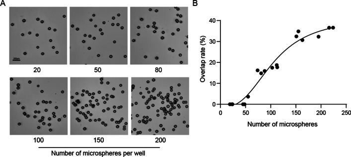
We have established a novel quantitative high-throughput imaging assay that harnesses the scalability of organoid cultures. Using this assay, we can capture organoid growth over time, measure multiple whole-well organoid readouts, and show the different responses to drug treatments.
In summary, combining the Z-stack imaging technique and fluorescent labeling methods, we established an assay for the imaging and analysis of three-dimensional organoids. Our data demonstrated the feasibility of using organoid-based platforms for high-throughput drug screening assays.
类器官是一种能够高度重现组织架构和细胞组成的三维结构,因此在基于类器官的药物筛选中具有广阔的应用前景。尽管在三维环境中生长为类器官重现相应组织的主要特征提供了可能性,但这使得在二维环境中对类器官进行成像以及在类器官受到辐射或化学疗法处理后,从周围死亡细胞中识别存活的类器官变得很不方便。因此,仍然需要开展大量工作来建立高质量的对照,以规范类器官分析并使类器官模型更具可重复性。
在这项研究中,我们使用 Z -stack 成像技术对三维类器官进行成像,以在一次成像中获取所有类器官的最大横截面。活细胞染色荧光染料 Calcein-AM 与 ImageJ 评估的结合用于分析辐射或化学疗法处理后的类器官的存活情况。
我们建立了一种新颖的定量高通量成像测定法,利用类器官培养的可扩展性。使用该测定法,我们可以捕获类器官随时间的生长情况,测量整个孔板的多个类器官读数,并展示对药物治疗的不同反应。
总之,我们结合 Z-stack 成像技术和荧光标记方法,建立了一种用于三维类器官成像和分析的测定法。我们的数据证明了基于类器官的平台在高通量药物筛选测定中的可行性。