• 文献检索
  • 文档翻译
  • 深度研究
  • 学术资讯
  • Suppr Zotero 插件Zotero 插件
  • 邀请有礼
  • 套餐&价格
  • 历史记录
应用&插件
Suppr Zotero 插件Zotero 插件浏览器插件Mac 客户端Windows 客户端微信小程序
定价
高级版会员购买积分包购买API积分包
服务
文献检索文档翻译深度研究API 文档MCP 服务
关于我们
关于 Suppr公司介绍联系我们用户协议隐私条款
关注我们

Suppr 超能文献

核心技术专利:CN118964589B侵权必究
粤ICP备2023148730 号-1Suppr @ 2026

文献检索

告别复杂PubMed语法,用中文像聊天一样搜索,搜遍4000万医学文献。AI智能推荐,让科研检索更轻松。

立即免费搜索

文件翻译

保留排版,准确专业,支持PDF/Word/PPT等文件格式,支持 12+语言互译。

免费翻译文档

深度研究

AI帮你快速写综述,25分钟生成高质量综述,智能提取关键信息,辅助科研写作。

立即免费体验

动态对比显微镜光学相干断层扫描作为一种评估角膜上皮暴露于苯扎氯铵时的新方法。

Dynamic Contrast Microscopic Optical Coherence Tomography As a Novel Method for Assessing Corneal Epithelium During Exposure to Benzalkonium Chloride.

机构信息

University Hospital Cologne, Cologne, Germany.

Medical Laser Center Lübeck GmbH, Lübeck, Germany.

出版信息

Transl Vis Sci Technol. 2022 May 2;11(5):28. doi: 10.1167/tvst.11.5.28.

DOI:10.1167/tvst.11.5.28
PMID:35622381
原文链接:https://pmc.ncbi.nlm.nih.gov/articles/PMC9145126/
Abstract

PURPOSE

Microscopic optical coherence tomography (mOCT) has an imaging resolution of 1 µm in all voxel dimensions, but individual epithelial cells are difficult to resolve due to lack of scattering contrast. Adding dynamic contrast processing to mOCT (dmOCT) results in color images that enable visualization of individual cells and possibly give information on cellular function via the calculation of a motility coefficient. We propose this technique as a novel method of evaluating the ocular surface after exposure to a toxic chemical, benzalkonium chloride (BAK).

METHODS

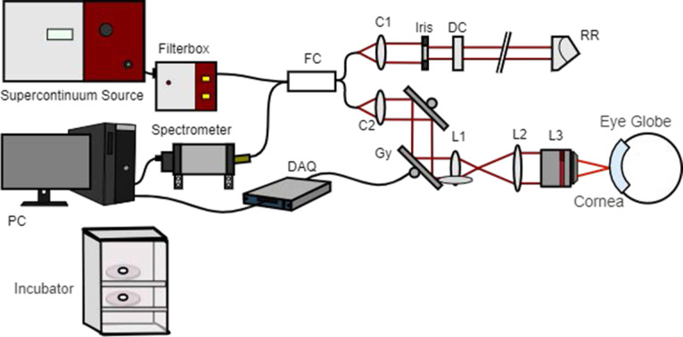
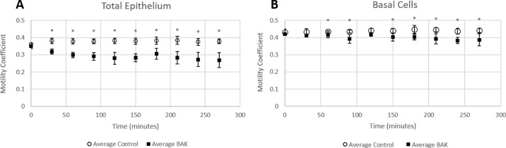
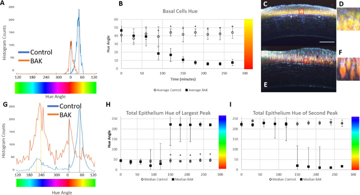
Ex vivo cross-section images were acquired with a custom-built, frequency-domain mOCT system. Eyes were explanted from healthy adult C57BL/6 mice and imaged every 30 minutes with five sets of dmOCT scans at each imaging time. Total epithelium and stroma thicknesses were measured from a single mOCT B-scan, and measures of color changes (hue) and the motility coefficient were acquired from dmOCT scans.

RESULTS

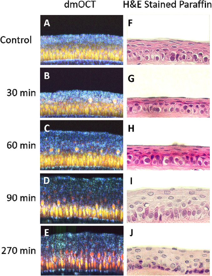
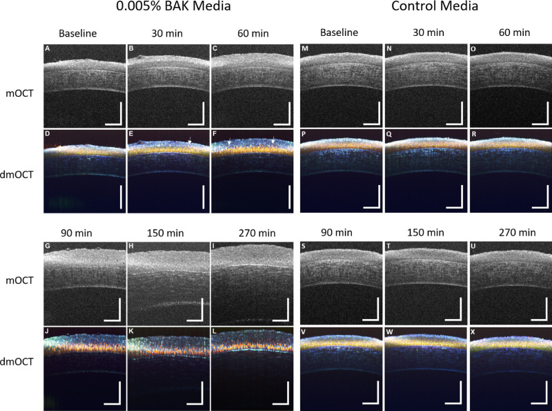
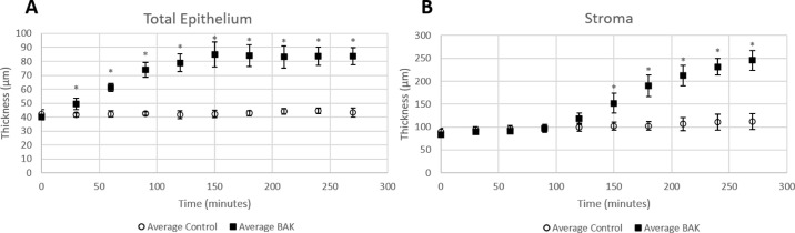
After 30-minute exposures to 0.005% BAK, local motility decreased and total epithelium thickness increased compared to controls. For basal epithelium cells, local motility decreased after 60-minute exposures, and the hue shifted red after 90-minute exposures. Stroma thickness did not significantly swell until 150-minute exposures to BAK.

CONCLUSIONS

dmOCT allows us to view the behavior of the cornea epithelium under toxic stress due to BAK, revealing parallel swelling of the extracellular matrix and changes in local subcellular motion.

TRANSLATIONAL RELEVANCE

The evaluation of the cornea epithelium using dmOCT is helpful to our understanding of the toxic effects of BAK.

摘要

目的

微观光学相干断层扫描(mOCT)在所有体素维度上的成像分辨率为 1 µm,但由于缺乏散射对比,单个上皮细胞难以分辨。在 mOCT(dmOCT)中添加动态对比处理会产生彩色图像,使单个细胞可视化,并通过计算运动系数提供有关细胞功能的信息。我们提出该技术作为评估暴露于有毒化学物质苯扎氯铵(BAK)后的眼表面的新方法。

方法

使用定制的频域 mOCT 系统获取离体横截面图像。从健康成年 C57BL/6 小鼠中取出眼睛,并每隔 30 分钟进行 5 组 dmOCT 扫描,每次成像时进行一次扫描。从单个 mOCT B 扫描中测量总上皮和基质厚度,并从 dmOCT 扫描中获取颜色变化(色调)和运动系数的测量值。

结果

暴露于 0.005% BAK 30 分钟后,与对照组相比,局部运动性降低,总上皮厚度增加。对于基底上皮细胞,暴露于 60 分钟后局部运动性降低,暴露于 90 分钟后色调变红。基质厚度直到暴露于 BAK 150 分钟后才明显肿胀。

结论

dmOCT 使我们能够观察到 BAK 引起的角膜上皮在有毒应激下的行为,揭示了细胞外基质的平行肿胀和局部亚细胞运动的变化。

翻译

田颖

https://cdn.ncbi.nlm.nih.gov/pmc/blobs/926c/9145126/4b8bc2b77e0f/tvst-11-5-28-f007.jpg
https://cdn.ncbi.nlm.nih.gov/pmc/blobs/926c/9145126/2b275c24c39c/tvst-11-5-28-f001.jpg
https://cdn.ncbi.nlm.nih.gov/pmc/blobs/926c/9145126/6fca97adbf76/tvst-11-5-28-f002.jpg
https://cdn.ncbi.nlm.nih.gov/pmc/blobs/926c/9145126/bada21a60aa5/tvst-11-5-28-f003.jpg
https://cdn.ncbi.nlm.nih.gov/pmc/blobs/926c/9145126/d33a39d4c29d/tvst-11-5-28-f004.jpg
https://cdn.ncbi.nlm.nih.gov/pmc/blobs/926c/9145126/e1a331daf234/tvst-11-5-28-f005.jpg
https://cdn.ncbi.nlm.nih.gov/pmc/blobs/926c/9145126/7776989a4747/tvst-11-5-28-f006.jpg
https://cdn.ncbi.nlm.nih.gov/pmc/blobs/926c/9145126/4b8bc2b77e0f/tvst-11-5-28-f007.jpg

相似文献

1
Dynamic Contrast Microscopic Optical Coherence Tomography As a Novel Method for Assessing Corneal Epithelium During Exposure to Benzalkonium Chloride.动态对比显微镜光学相干断层扫描作为一种评估角膜上皮暴露于苯扎氯铵时的新方法。
Transl Vis Sci Technol. 2022 May 2;11(5):28. doi: 10.1167/tvst.11.5.28.
2
Mapping of Corneal Layer Thicknesses With Polarization-Sensitive Optical Coherence Tomography Using a Conical Scan Pattern.利用锥形扫描模式的偏振敏感光学相干断层扫描对角膜层厚度进行映射。
Invest Ophthalmol Vis Sci. 2018 Nov 1;59(13):5579-5588. doi: 10.1167/iovs.18-25311.
3
The repeatability of corneal and corneal epithelial thickness measurements using optical coherence tomography.使用光学相干断层扫描技术测量角膜及角膜上皮厚度的可重复性。
Optom Vis Sci. 2006 Jun;83(6):360-5. doi: 10.1097/01.opx.0000221388.26031.23.
4
Benzalkonium chloride-induced direct and indirect toxicity on corneal epithelial and trigeminal neuronal cells: proinflammatory and apoptotic responses in vitro.苯扎氯铵对角膜上皮细胞和三叉神经神经元细胞的直接和间接毒性:体外的促炎和凋亡反应。
Toxicol Lett. 2020 Feb 1;319:74-84. doi: 10.1016/j.toxlet.2019.10.014. Epub 2019 Nov 7.
5
Evaluation of corneal epithelial thickness in glaucomatous patients using anterior-segment optical coherence tomography.应用眼前节光学相干断层扫描仪评估青光眼患者的角膜上皮厚度。
J Biophotonics. 2020 Jan;13(1):e201900095. doi: 10.1002/jbio.201900095. Epub 2019 Oct 29.
6
Label-Free In Vivo Imaging of Corneal Lymphatic Vessels Using Microscopic Optical Coherence Tomography.使用显微光学相干断层扫描技术对角膜淋巴管进行无标记活体成像。
Invest Ophthalmol Vis Sci. 2017 Nov 1;58(13):5880-5886. doi: 10.1167/iovs.17-22286.
7
Confocal microscopic studies of living rabbit cornea treated with benzalkonium chloride.用苯扎氯铵处理的活体兔角膜的共聚焦显微镜研究。
Cornea. 1992 May;11(3):221-5.
8
Microscopic optical coherence tomography (mOCT) at 600 kHz for 4D volumetric imaging and dynamic contrast.用于4D体积成像和动态造影的600千赫兹显微光学相干断层扫描(mOCT)
Biomed Opt Express. 2021 Sep 7;12(10):6024-6039. doi: 10.1364/BOE.425001. eCollection 2021 Oct 1.
9
Arc-scanning very high-frequency digital ultrasound for 3D pachymetric mapping of the corneal epithelium and stroma in laser in situ keratomileusis.用于准分子原位角膜磨镶术中角膜上皮和基质三维测厚成像的弧形扫描甚高频数字超声检查
J Refract Surg. 2000 Jul-Aug;16(4):414-30. doi: 10.3928/1081-597X-20000701-04.
10
Reliability and reproducibility of assessment of corneal epithelial thickness by fourier domain optical coherence tomography.傅里叶域光学相干断层扫描评估角膜上皮厚度的可靠性和可重复性。
Invest Ophthalmol Vis Sci. 2012 May 4;53(6):2580-5. doi: 10.1167/iovs.11-8981.

引用本文的文献

1
Fresnel biprism common-path low-coherence digital holography for dynamic light scattering spectroscopy of biological materials.用于生物材料动态光散射光谱分析的菲涅耳双棱镜共光路低相干数字全息术。
Biomed Opt Express. 2025 Jan 30;16(2):806-820. doi: 10.1364/BOE.543308. eCollection 2025 Feb 1.
2
Non-destructive viability assessment of cancer cell spheroids using dynamic optical coherence tomography with trypan blue validation.使用动态光学相干断层扫描并经台盼蓝验证对癌细胞球体进行非破坏性活力评估。
Biomed Opt Express. 2024 Oct 14;15(11):6370-6383. doi: 10.1364/BOE.533339. eCollection 2024 Nov 1.
3
Line-field dynamic optical coherence tomography platform for volumetric assessment of biological tissues.

本文引用的文献

1
Microscopic optical coherence tomography (mOCT) at 600 kHz for 4D volumetric imaging and dynamic contrast.用于4D体积成像和动态造影的600千赫兹显微光学相干断层扫描(mOCT)
Biomed Opt Express. 2021 Sep 7;12(10):6024-6039. doi: 10.1364/BOE.425001. eCollection 2021 Oct 1.
2
Benzalkonium Chloride-Preserved Anti-Glaucomatous Eye Drops and Their Effect on Human Conjunctival Goblet Cells in vitro.苯扎氯铵保存的抗青光眼滴眼液及其对人结膜杯状细胞的体外作用。
Biomed Hub. 2021 Aug 13;6(2):69-75. doi: 10.1159/000517845. eCollection 2021 May-Aug.
3
Changes in Corneal Epithelial Thickness Induced by Topical Antiglaucoma Medications.
用于生物组织体积评估的线场动态光学相干断层扫描平台。
Biomed Opt Express. 2024 Jun 7;15(7):4162-4175. doi: 10.1364/BOE.527797. eCollection 2024 Jul 1.
4
Cellular structural and functional imaging of donor and pathological corneas with label-free dual-mode full-field optical coherence tomography.使用无标记双模式全场光学相干断层扫描对供体角膜和病理角膜进行细胞结构和功能成像。
Biomed Opt Express. 2024 May 21;15(6):3869-3888. doi: 10.1364/BOE.525116. eCollection 2024 Jun 1.
5
Label-free drug response evaluation of human derived tumor spheroids using three-dimensional dynamic optical coherence tomography.利用三维动态光学相干断层成像技术对人源肿瘤球体进行无标记药物反应评估。
Sci Rep. 2023 Sep 16;13(1):15377. doi: 10.1038/s41598-023-41846-3.
6
Dynamic optical coherence tomography for cell analysis [Invited].用于细胞分析的动态光学相干断层扫描[特邀报告]
Biomed Opt Express. 2023 Jun 14;14(7):3362-3379. doi: 10.1364/BOE.488929. eCollection 2023 Jul 1.
局部抗青光眼药物引起的角膜上皮厚度变化
J Clin Med. 2021 Aug 5;10(16):3464. doi: 10.3390/jcm10163464.
4
Ocular Surface - Merging Challenges and Opportunities.眼表——融合挑战与机遇
Transl Vis Sci Technol. 2020 Nov 2;9(12):3. doi: 10.1167/tvst.9.12.3. eCollection 2020 Nov.
5
Micro-optical coherence tomography for high-resolution morphologic imaging of cellular and nerval corneal micro-structures.用于细胞和神经角膜微结构高分辨率形态学成像的显微光学相干断层扫描技术
Biomed Opt Express. 2020 Sep 28;11(10):5920-5933. doi: 10.1364/BOE.402971. eCollection 2020 Oct 1.
6
Dynamic contrast in scanning microscopic OCT.扫描显微镜光学相干断层扫描中的动态对比度。
Opt Lett. 2020 Sep 1;45(17):4766-4769. doi: 10.1364/OL.396134.
7
Dynamic full-field optical coherence tomography: 3D live-imaging of retinal organoids.动态全场光学相干断层扫描:视网膜类器官的三维实时成像
Light Sci Appl. 2020 Aug 17;9:140. doi: 10.1038/s41377-020-00375-8. eCollection 2020.
8
Stromal Nerve Imaging and Tracking Using Micro-Optical Coherence Tomography.基质神经成像和跟踪使用微光学相干断层扫描。
Transl Vis Sci Technol. 2020 Apr 15;9(5):6. doi: 10.1167/tvst.9.5.6. eCollection 2020 Apr.
9
Imaging intracellular motion with dynamic micro-optical coherence tomography.利用动态微光学相干断层扫描成像技术对细胞内运动进行成像。
Biomed Opt Express. 2020 Apr 28;11(5):2768-2778. doi: 10.1364/BOE.390782. eCollection 2020 May 1.
10
Real-time non-contact cellular imaging and angiography of human cornea and limbus with common-path full-field/SD OCT.应用共路全视野/SD-OCT 对人眼角膜和角膜缘进行实时非接触式细胞成像和血管造影。
Nat Commun. 2020 Apr 20;11(1):1868. doi: 10.1038/s41467-020-15792-x.