School of Molecular Sciences, Biodesign Center for Applied Structural Discovery, Arizona State University, Tempe, AZ 85287, USA.
Biomolecules. 2022 Apr 24;12(5):628. doi: 10.3390/biom12050628.
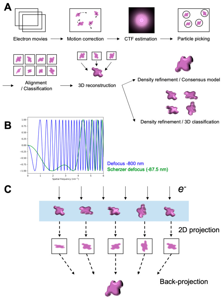
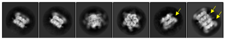
Single-particle cryogenic electron microscopy (cryo-EM) has become an indispensable tool to probe high-resolution structural detail of biomolecules. It enables direct visualization of the biomolecules and opens a possibility for averaging molecular images to reconstruct a three-dimensional Coulomb potential density map. Newly developed algorithms for data analysis allow for the extraction of structural heterogeneity from a massive and low signal-to-noise-ratio (SNR) cryo-EM dataset, expanding our understanding of multiple conformational states, or further implications in dynamics, of the target biomolecule. This review provides an overview that briefly describes the workflow of single-particle cryo-EM, including imaging and data processing, and new methods developed for analyzing the data heterogeneity to understand the structural variability of biomolecules.
单颗粒低温电子显微镜(cryo-EM)已成为探测生物分子高分辨率结构细节的不可或缺的工具。它能够直接观察生物分子,并为平均分子图像以重建三维库仑位密度图提供了可能性。新开发的数据分析算法允许从大量低信噪比(SNR)的 cryo-EM 数据集中提取结构异质性,从而扩展我们对目标生物分子的多种构象状态或动态的理解。本文综述简要描述了单颗粒 cryo-EM 的工作流程,包括成像和数据处理,以及为分析数据异质性以了解生物分子结构可变性而开发的新方法。