Granata Vincenza, Fusco Roberta, De Muzio Federica, Cutolo Carmen, Mattace Raso Mauro, Gabelloni Michela, Avallone Antonio, Ottaiano Alessandro, Tatangelo Fabiana, Brunese Maria Chiara, Miele Vittorio, Izzo Francesco, Petrillo Antonella
Division of Radiology, Istituto Nazionale Tumori IRCCS Fondazione Pascale-IRCCS di Napoli, 80131 Naples, Italy.
Medical Oncology Division, Igea SpA, 41012 Carpi, Italy.
Diagnostics (Basel). 2022 Apr 29;12(5):1115. doi: 10.3390/diagnostics12051115.
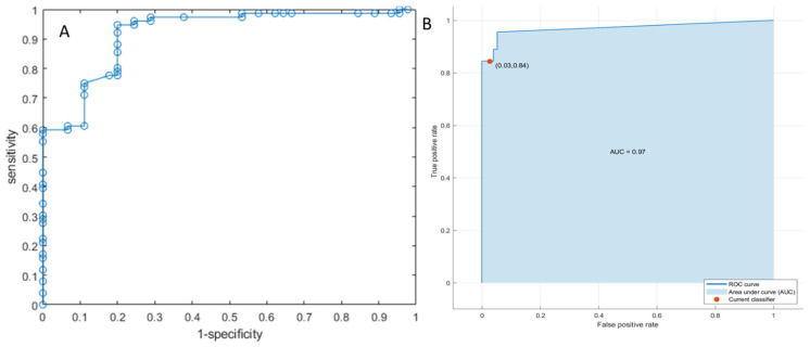
To assess Radiomics and Machine Learning Analysis in Liver Colon and Rectal Cancer Metastases (CRLM) Growth Pattern, we evaluated, retrospectively, a training set of 51 patients with 121 liver metastases and an external validation set of 30 patients with a single lesion. All patients were subjected to MRI studies in pre-surgical setting. For each segmented volume of interest (VOI), 851 radiomics features were extracted using PyRadiomics package. Nonparametric test, univariate, linear regression analysis and patter recognition approaches were performed. The best results to discriminate expansive versus infiltrative front of tumor growth with the highest accuracy and AUC at univariate analysis were obtained by the wavelet_LHH_glrlm_ShortRunLowGray Level Emphasis from portal phase of contrast study. With regard to linear regression model, this increased the performance obtained respect to the univariate analysis for each sequence except that for EOB-phase sequence. The best results were obtained by a linear regression model of 15 significant features extracted by the T2-W SPACE sequence. Furthermore, using pattern recognition approaches, the diagnostic performance to discriminate the expansive versus infiltrative front of tumor growth increased again and the best classifier was a weighted KNN trained with the 9 significant metrics extracted from the portal phase of contrast study, with an accuracy of 92% on training set and of 91% on validation set. In the present study, we have demonstrated as Radiomics and Machine Learning Analysis, based on EOB-MRI study, allow to identify several biomarkers that permit to recognise the different Growth Patterns in CRLM.
为了评估肝脏结直肠癌转移(CRLM)生长模式的影像组学和机器学习分析,我们回顾性评估了一个包含51例患者共121个肝转移灶的训练集,以及一个包含30例患者单个病灶的外部验证集。所有患者在术前均接受了MRI检查。对于每个分割的感兴趣体积(VOI),使用PyRadiomics软件包提取了851个影像组学特征。进行了非参数检验、单变量分析、线性回归分析和模式识别方法。在单变量分析中,通过对比研究门静脉期的小波_LHH_glrlm_ShortRunLowGray Level Emphasis获得了区分肿瘤生长的扩张性与浸润性前沿的最佳结果,其具有最高的准确性和AUC。对于线性回归模型,除了EOB期序列外,每个序列相对于单变量分析的性能都有所提高。通过T2-W SPACE序列提取的15个显著特征的线性回归模型获得了最佳结果。此外,使用模式识别方法,区分肿瘤生长的扩张性与浸润性前沿的诊断性能再次提高,最佳分类器是使用从对比研究门静脉期提取的9个显著指标训练的加权KNN,在训练集上的准确率为92%,在验证集上的准确率为91%。在本研究中,我们已经证明基于EOB-MRI研究的影像组学和机器学习分析能够识别几种生物标志物,这些生物标志物有助于识别CRLM中的不同生长模式。