Park Sun Mi, Kim Jisu, Baek Suji, Jeon Joo-Yeong, Lee Sang Ju, Kang Seo Young, Yoo Min Young, Yoon Hai-Jeon, Kwon Seung Hae, Lim Kiwon, Oh Seung Jun, Kim Bom Sahn, Lee Kang Pa, Moon Byung Seok
Department of Nuclear Medicine, College of Medicine, Ewha Womans University Seoul Hospital, Ewha Womans University, Seoul 07804, Korea.
Physical Activity and Performance Institute, Konkuk University, Seoul 05029, Korea.
Diagnostics (Basel). 2022 May 20;12(5):1274. doi: 10.3390/diagnostics12051274.
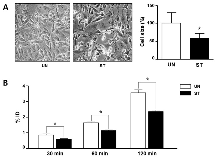
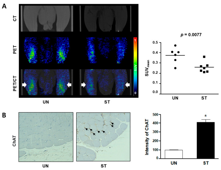
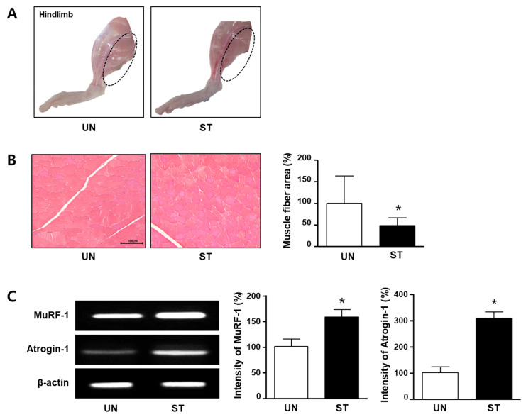
Imaging techniques for diagnosing muscle atrophy and sarcopenia remain insufficient, although various advanced diagnostic methods have been established. We explored the feasibility of F-fluorocholine (F-FCH) positron emission tomography/computed tomography (PET/CT) for evaluating skeletal muscle atrophy, as an imaging technique that tracks choline level changes in muscles. Cell uptake in L6 cells by F-FCH was performed in a complete medium containing serum (untreated group, UN) and a serum-free medium (starved group, ST). Small-animal-dedicated PET/CT imaging with F-FCH was examined in in-vivo models with rats that were starved for 2 days to cause muscle atrophy. After the hind limbs were dissected, starvation-induced in-vivo models were anatomically confirmed by reverse-transcription polymerase chain reaction to evaluate the expression levels of the atrophy markers muscle RING-finger protein-1 (MuRF-1) and atrogin-1. F-FCH uptake was lower in the starvation-induced cells than in the untreated group, and in-vivo PET uptake also revealed a similar tendency (the average standardized uptake value (SUV) = 0.26 ± 0.06 versus 0.37 ± 0.07, respectively). Furthermore, the expression levels of MuRF-1 and atrogin-1 mRNA were significantly increased in the starvation-induced muscle atrophy of rats compared to the untreated group. F-FCH PET/CT may be a promising tool for diagnosing skeletal muscle atrophy.
尽管已经建立了各种先进的诊断方法,但用于诊断肌肉萎缩和肌少症的成像技术仍然不足。我们探索了F-氟胆碱(F-FCH)正电子发射断层扫描/计算机断层扫描(PET/CT)作为一种追踪肌肉中胆碱水平变化的成像技术来评估骨骼肌萎缩的可行性。在含有血清的完全培养基(未处理组,UN)和无血清培养基(饥饿组,ST)中进行F-FCH对L6细胞的摄取。在饥饿2天导致肌肉萎缩的大鼠体内模型中检查了F-FCH的小动物专用PET/CT成像。解剖后肢后,通过逆转录聚合酶链反应对饥饿诱导的体内模型进行解剖学确认,以评估萎缩标志物肌肉环指蛋白-1(MuRF-1)和atrogin-1的表达水平。饥饿诱导的细胞中F-FCH摄取低于未处理组,体内PET摄取也显示出类似趋势(平均标准化摄取值(SUV)分别为0.26±0.06和0.37±0.07)。此外,与未处理组相比,饥饿诱导的大鼠肌肉萎缩中MuRF-1和atrogin-1 mRNA的表达水平显著增加。F-FCH PET/CT可能是诊断骨骼肌萎缩的一种有前途的工具。