Oyibo Prosper, Jujjavarapu Satyajith, Meulah Brice, Agbana Tope, Braakman Ingeborg, van Diepen Angela, Bengtson Michel, van Lieshout Lisette, Oyibo Wellington, Vdovine Gleb, Diehl Jan-Carel
Delft Center for Systems and Control, Faculty of Mechanical, Maritime and Materials Engineering, Delft University of Technology, 2628 CD Delft, The Netherlands.
ANDI Centre of Excellence for Malaria Diagnosis, College of Medicine, University of Lagos, Lagos 101017, Nigeria.
Micromachines (Basel). 2022 Apr 19;13(5):643. doi: 10.3390/mi13050643.
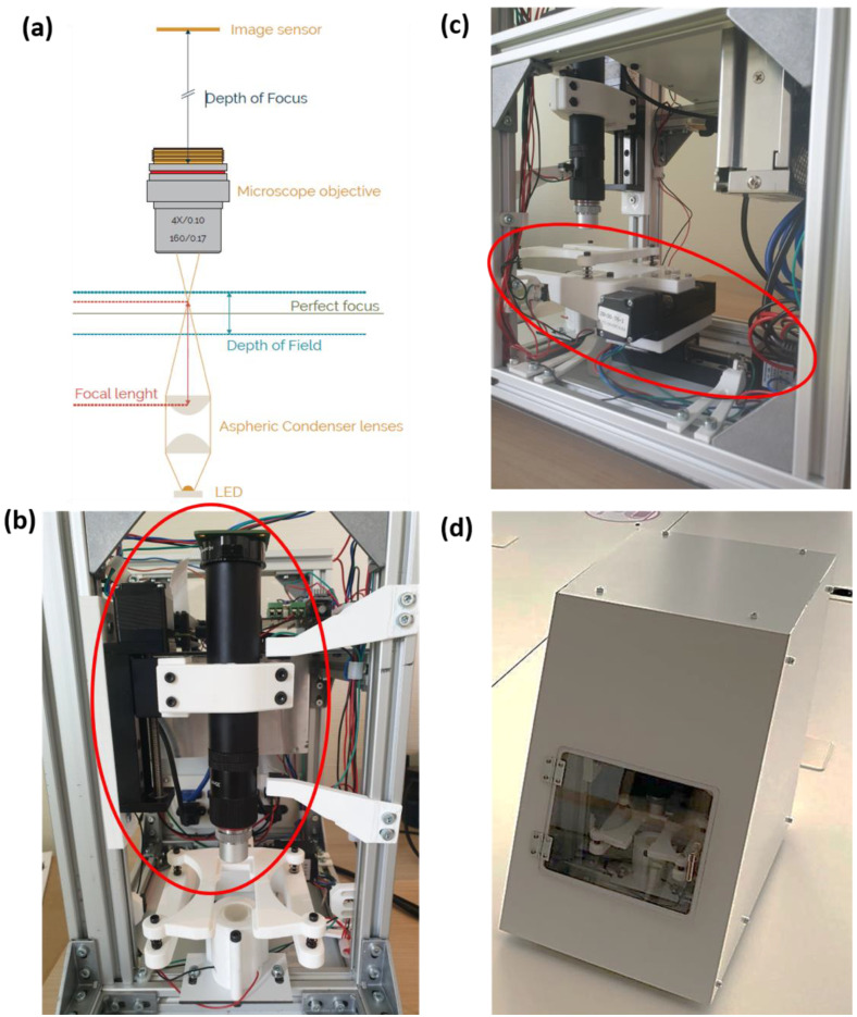
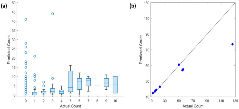
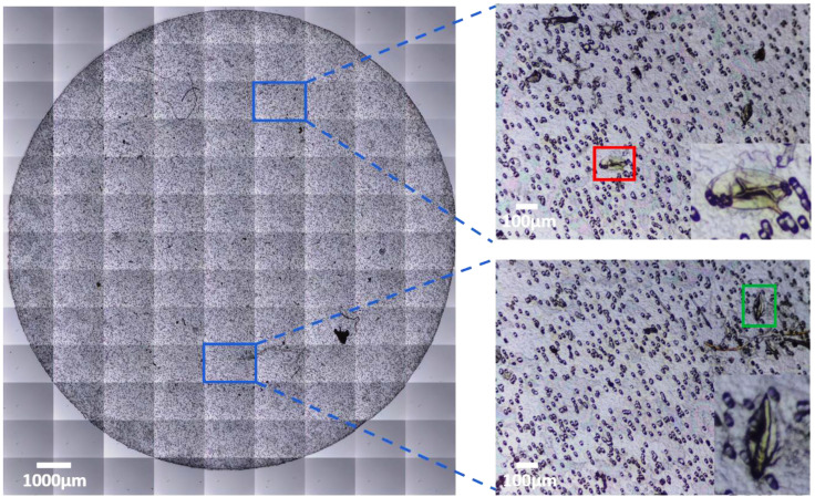
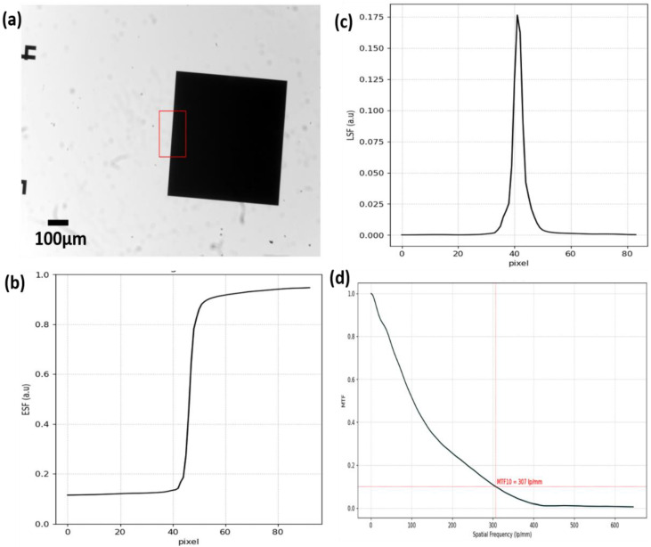
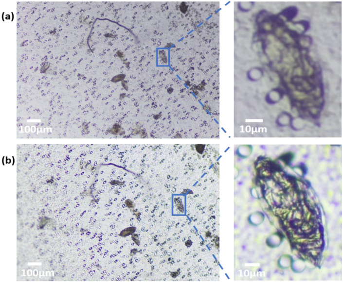
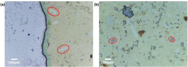
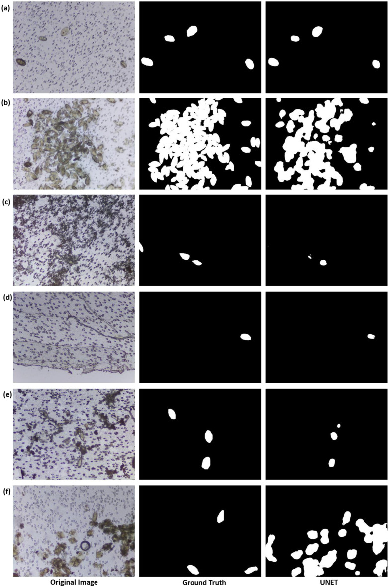
For many parasitic diseases, the microscopic examination of clinical samples such as urine and stool still serves as the diagnostic reference standard, primarily because microscopes are accessible and cost-effective. However, conventional microscopy is laborious, requires highly skilled personnel, and is highly subjective. Requirements for skilled operators, coupled with the cost and maintenance needs of the microscopes, which is hardly done in endemic countries, presents grossly limited access to the diagnosis of parasitic diseases in resource-limited settings. The urgent requirement for the management of tropical diseases such as schistosomiasis, which is now focused on elimination, has underscored the critical need for the creation of access to easy-to-use diagnosis for case detection, community mapping, and surveillance. In this paper, we present a low-cost automated digital microscope-the Schistoscope-which is capable of automatic focusing and scanning regions of interest in prepared microscope slides, and automatic detection of eggs in captured images. The device was developed using widely accessible distributed manufacturing methods and off-the-shelf components to enable local manufacturability and ease of maintenance. For proof of principle, we created a egg dataset of over 5000 images captured from spiked and clinical urine samples from field settings and demonstrated the automatic detection of eggs using a trained deep neural network model. The experiments and results presented in this paper collectively illustrate the robustness, stability, and optical performance of the device, making it suitable for use in the monitoring and evaluation of schistosomiasis control programs in endemic settings.
对于许多寄生虫病而言,对尿液和粪便等临床样本进行显微镜检查仍然是诊断的参考标准,主要原因是显微镜易于获取且成本效益高。然而,传统显微镜检查工作繁琐,需要高技能人员操作,且主观性很强。对熟练操作人员的要求,再加上显微镜的成本和维护需求,在流行国家很难做到,这使得在资源有限的环境中,寄生虫病诊断的可及性极为有限。对血吸虫病等热带疾病管理的迫切需求,目前重点在于消除该疾病,这凸显了创建易于使用的诊断方法以进行病例检测、社区绘图和监测的迫切需要。在本文中,我们展示了一种低成本的自动化数字显微镜——血吸虫镜,它能够自动聚焦和扫描制备好的显微镜载玻片上的感兴趣区域,并能自动检测捕获图像中的虫卵。该设备是使用广泛可用的分布式制造方法和现成组件开发的,以实现本地制造能力和易于维护。为了验证原理,我们创建了一个包含5000多张图像的虫卵数据集,这些图像是从现场采集的加标和临床尿液样本中捕获的,并使用经过训练的深度神经网络模型展示了虫卵的自动检测。本文展示的实验和结果共同说明了该设备的稳健性、稳定性和光学性能,使其适用于流行地区血吸虫病控制项目的监测和评估。