Nash Family Department of Neuroscience, Icahn School of Medicine at Mount Sinai, New York, United States.
Department of Neurology, David Geffen School of Medicine, University of California, Los Angeles, Los Angeles, United States.
Elife. 2022 Jun 1;11:e70661. doi: 10.7554/eLife.70661.
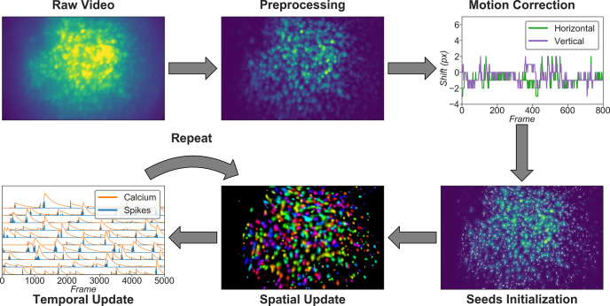
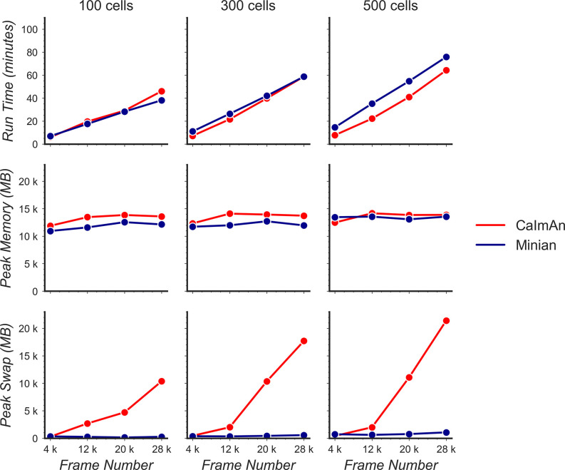
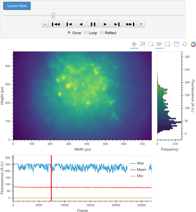
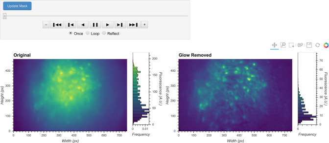
Miniature microscopes have gained considerable traction for in vivo calcium imaging in freely behaving animals. However, extracting calcium signals from raw videos is a computationally complex problem and remains a bottleneck for many researchers utilizing single-photon in vivo calcium imaging. Despite the existence of many powerful analysis packages designed to detect and extract calcium dynamics, most have either key parameters that are hard-coded or insufficient step-by-step guidance and validations to help the users choose the best parameters. This makes it difficult to know whether the output is reliable and meets the assumptions necessary for proper analysis. Moreover, large memory demand is often a constraint for setting up these pipelines since it limits the choice of hardware to specialized computers. Given these difficulties, there is a need for a low memory demand, user-friendly tool offering interactive visualizations of how altering parameters at each step of the analysis affects data output. Our open-source analysis pipeline, Minian (miniscope analysis), facilitates the transparency and accessibility of single-photon calcium imaging analysis, permitting users with little computational experience to extract the location of cells and their corresponding calcium traces and deconvolved neural activities. Minian contains interactive visualization tools for every step of the analysis, as well as detailed documentation and tips on parameter exploration. Furthermore, Minian has relatively small memory demands and can be run on a laptop, making it available to labs that do not have access to specialized computational hardware. Minian has been validated to reliably and robustly extract calcium events across different brain regions and from different cell types. In practice, Minian provides an open-source calcium imaging analysis pipeline with user-friendly interactive visualizations to explore parameters and validate results.
微型显微镜在自由活动动物的体内钙成像中得到了广泛的关注。然而,从原始视频中提取钙信号是一个计算复杂的问题,仍然是许多利用单光子体内钙成像的研究人员的一个瓶颈。尽管有许多强大的分析软件包旨在检测和提取钙动力学,但大多数软件包要么关键参数是硬编码的,要么缺乏逐步的指导和验证,以帮助用户选择最佳参数。这使得很难知道输出是否可靠,并且是否满足适当分析所需的假设。此外,由于设置这些管道需要大量的内存,因此通常会受到限制,因为它限制了硬件的选择只能使用专门的计算机。鉴于这些困难,需要一种低内存需求、用户友好的工具,提供如何在分析的每个步骤更改参数会如何影响数据输出的交互式可视化。我们的开源分析管道 Minian(微型显微镜分析)促进了单光子钙成像分析的透明度和可访问性,允许具有较少计算经验的用户提取细胞的位置及其相应的钙迹线和去卷积神经活动。Minian 包含分析每个步骤的交互式可视化工具,以及关于参数探索的详细文档和提示。此外,Minian 的内存需求相对较小,可以在笔记本电脑上运行,这使得没有专用计算硬件的实验室也可以使用它。Minian 已被验证能够可靠和稳健地提取不同脑区和不同细胞类型的钙事件。在实践中,Minian 提供了一个具有用户友好的交互式可视化功能的开源钙成像分析管道,用于探索参数和验证结果。