Department of Ophthalmology, University Hospital Bonn, Ernst-Abbe-Str. 2, 53127, Bonn, Germany.
Department of Neurology, University Hospital Bonn, Bonn, Germany.
Sci Rep. 2022 Jun 3;12(1):9315. doi: 10.1038/s41598-022-13312-z.
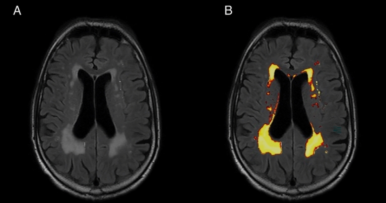
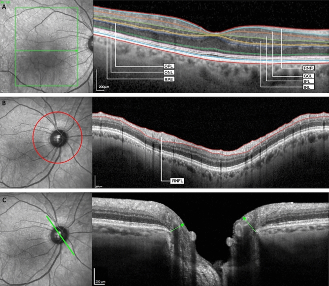
Cerebral small vessel disease (CSVD) is an important contributor to cognitive impairment and stroke. Previous research has suggested associations with alterations in single retinal layers. We have assessed changes of all individual retinal layers in CSVD using high-resolution optical coherence tomography (OCT) for the first time. Subjects with recent magnetic resonance imaging (MRI) underwent macular and peripapillary retinal imaging using OCT for this case-control study. Number and volume ratio index (WMRI) of white matter lesions (WML) were determined on MRI. Data were analyzed using multiple linear regression models. 27 CSVD patients and 9 control participants were included. Ganglion cell layer (GCL) volume was significantly reduced in patients with CSVD compared to age-matched controls (p = 0.008). In patients with CSVD, larger foveal outer plexiform layer (OPL) volume and decreased temporal peripapillary retinal nerve fiber layer (RNFL) thickness were significantly associated with a higher WMRI in linear regression when controlling for age (p ≤ 0.033). Decreased foveal GCL volume and temporal-inferior RNFL thickness at Bruch's membrane opening (MRW), and increased temporal MRW were associated with a higher WML burden (p ≤ 0.037). Thus, we identified alterations in several OCT layers in individuals with CSVD (GCL, OPL, MRW and RNFL). Their potential diagnostic value merits further study.
脑小血管病(CSVD)是认知障碍和中风的重要原因。先前的研究表明,它与单一视网膜层的改变有关。我们首次使用高分辨率光学相干断层扫描(OCT)评估了 CSVD 中所有单个视网膜层的变化。这项病例对照研究中,有近期磁共振成像(MRI)的受试者使用 OCT 进行了黄斑和视盘周围视网膜成像。在 MRI 上确定了脑白质病变(WML)的数量和体积比指数(WMRI)。使用多元线性回归模型对数据进行分析。共纳入 27 例 CSVD 患者和 9 例对照参与者。与年龄匹配的对照组相比,CSVD 患者的神经节细胞层(GCL)体积明显减小(p=0.008)。在 CSVD 患者中,当控制年龄时,较大的中心凹外丛状层(OPL)体积和颞侧视盘周围视网膜神经纤维层(RNFL)厚度减小与较高的 WMRI 呈线性相关(p≤0.033)。在布吕赫膜开口处(MRW)的神经节细胞层(GCL)体积减小、颞下 RNFL 厚度减小和颞侧 MRW 增大与较高的 WML 负担相关(p≤0.037)。因此,我们在 CSVD 患者中发现了几个 OCT 层的改变(GCL、OPL、MRW 和 RNFL)。它们的潜在诊断价值值得进一步研究。