• 文献检索
  • 文档翻译
  • 深度研究
  • 学术资讯
  • Suppr Zotero 插件Zotero 插件
  • 邀请有礼
  • 套餐&价格
  • 历史记录
应用&插件
Suppr Zotero 插件Zotero 插件浏览器插件Mac 客户端Windows 客户端微信小程序
定价
高级版会员购买积分包购买API积分包
服务
文献检索文档翻译深度研究API 文档MCP 服务
关于我们
关于 Suppr公司介绍联系我们用户协议隐私条款
关注我们

Suppr 超能文献

核心技术专利:CN118964589B侵权必究
粤ICP备2023148730 号-1Suppr @ 2026

文献检索

告别复杂PubMed语法,用中文像聊天一样搜索,搜遍4000万医学文献。AI智能推荐,让科研检索更轻松。

立即免费搜索

文件翻译

保留排版,准确专业,支持PDF/Word/PPT等文件格式,支持 12+语言互译。

免费翻译文档

深度研究

AI帮你快速写综述,25分钟生成高质量综述,智能提取关键信息,辅助科研写作。

立即免费体验

相似文献

1
Learning Semantics-enriched Representation via Self-discovery, Self-classification, and Self-restoration.通过自我发现、自我分类和自我恢复学习语义丰富的表示。
Med Image Comput Comput Assist Interv. 2020 Oct;12261:137-147. doi: 10.1007/978-3-030-59710-8_14. Epub 2020 Sep 29.
2
Models Genesis.模型起源。
Med Image Anal. 2021 Jan;67:101840. doi: 10.1016/j.media.2020.101840. Epub 2020 Oct 13.
3
Models Genesis: Generic Autodidactic Models for 3D Medical Image Analysis.模型起源:用于3D医学图像分析的通用自学习模型
Med Image Comput Comput Assist Interv. 2019 Oct;11767:384-393. doi: 10.1007/978-3-030-32251-9_42. Epub 2019 Oct 10.
4
Transferable Visual Words: Exploiting the Semantics of Anatomical Patterns for Self-Supervised Learning.可转移的视觉词汇:利用解剖模式的语义进行自监督学习。
IEEE Trans Med Imaging. 2021 Oct;40(10):2857-2868. doi: 10.1109/TMI.2021.3060634. Epub 2021 Sep 30.
5
Self-supervised learning for medical image analysis: Discriminative, restorative, or adversarial?用于医学图像分析的自监督学习:判别式、恢复式还是对抗式?
Med Image Anal. 2024 May;94:103086. doi: 10.1016/j.media.2024.103086. Epub 2024 Jan 28.
6
Linear semantic transformation for semi-supervised medical image segmentation.线性语义变换在半监督医学图像分割中的应用。
Comput Biol Med. 2024 May;173:108331. doi: 10.1016/j.compbiomed.2024.108331. Epub 2024 Mar 21.
7
A Systematic Benchmarking Analysis of Transfer Learning for Medical Image Analysis.医学图像分析中迁移学习的系统基准分析
Domain Adapt Represent Transf Afford Healthc AI Resour Divers Glob Health (2021). 2021 Sep-Oct;12968:3-13. doi: 10.1007/978-3-030-87722-4_1. Epub 2021 Sep 21.
8
DiRA: Discriminative, Restorative, and Adversarial Learning for Self-supervised Medical Image Analysis.DiRA:用于自监督医学图像分析的判别式、恢复式和对抗式学习
Proc IEEE Comput Soc Conf Comput Vis Pattern Recognit. 2022 Jun;2022:20792-20802. doi: 10.1109/cvpr52688.2022.02016. Epub 2022 Sep 27.
9
Group-Wise Learning for Weakly Supervised Semantic Segmentation.基于群体学习的弱监督语义分割。
IEEE Trans Image Process. 2022;31:799-811. doi: 10.1109/TIP.2021.3132834. Epub 2022 Jan 4.
10
Parts2Whole: Self-supervised Contrastive Learning via Reconstruction.从部分到整体:通过重建进行自监督对比学习
Domain Adapt Represent Transf Distrib Collab Learn (2020). 2020 Oct;12444:85-95. doi: 10.1007/978-3-030-60548-3_9. Epub 2020 Sep 26.

引用本文的文献

1
Representing Part-Whole Hierarchies in Foundation Models by Learning Localizability, Composability, and Decomposability from Anatomy via Self-Supervision.通过自监督从解剖学中学习局部可定位性、可组合性和可分解性,在基础模型中表示部分-整体层次结构。
Proc IEEE Comput Soc Conf Comput Vis Pattern Recognit. 2024 Jun;abs/210504906(2024):11269-11281. doi: 10.1109/cvpr52733.2024.01071. Epub 2024 Sep 16.
2
Advancements and Challenges in the Image-Based Diagnosis of Lung and Colon Cancer: A Comprehensive Review.肺癌和结肠癌基于图像诊断的进展与挑战:综述
Cancer Inform. 2024 Oct 16;23:11769351241290608. doi: 10.1177/11769351241290608. eCollection 2024.
3
Self-supervised learning framework application for medical image analysis: a review and summary.基于自监督学习框架的医学图像分析应用:综述与总结。
Biomed Eng Online. 2024 Oct 27;23(1):107. doi: 10.1186/s12938-024-01299-9.
4
Selection of pre-trained weights for transfer learning in automated cytomegalovirus retinitis classification.用于自动化巨细胞病毒视网膜炎分类的迁移学习中预训练权重的选择。
Sci Rep. 2024 Jul 10;14(1):15899. doi: 10.1038/s41598-024-67121-7.
5
A survey of the impact of self-supervised pretraining for diagnostic tasks in medical X-ray, CT, MRI, and ultrasound.针对医学 X 射线、CT、MRI 和超声诊断任务的自监督预训练的影响进行调查。
BMC Med Imaging. 2024 Apr 6;24(1):79. doi: 10.1186/s12880-024-01253-0.
6
Self-supervised learning for medical image analysis: Discriminative, restorative, or adversarial?用于医学图像分析的自监督学习:判别式、恢复式还是对抗式?
Med Image Anal. 2024 May;94:103086. doi: 10.1016/j.media.2024.103086. Epub 2024 Jan 28.
7
Decoding phenotypic screening: A comparative analysis of image representations.解码表型筛选:图像表示的比较分析
Comput Struct Biotechnol J. 2024 Mar 12;23:1181-1188. doi: 10.1016/j.csbj.2024.02.022. eCollection 2024 Dec.
8
Improving Computer-Aided Thoracic Disease Diagnosis through Comparative Analysis Using Chest X-ray Images Taken at Different Times.通过对不同时间拍摄的胸部 X 光图像进行比较分析,提高计算机辅助胸部疾病诊断水平。
Sensors (Basel). 2024 Feb 24;24(5):1478. doi: 10.3390/s24051478.
9
Seeking an optimal approach for Computer-aided Diagnosis of Pulmonary Embolism.寻求肺栓塞计算机辅助诊断的最佳方法。
Med Image Anal. 2024 Jan;91:102988. doi: 10.1016/j.media.2023.102988. Epub 2023 Oct 13.
10
Anatomy-Aware Contrastive Representation Learning for Fetal Ultrasound.用于胎儿超声的解剖学感知对比表示学习
Comput Vis ECCV. 2022 Oct;2022:422-436. doi: 10.1007/978-3-031-25066-8_23.

本文引用的文献

1
Models Genesis: Generic Autodidactic Models for 3D Medical Image Analysis.模型起源:用于3D医学图像分析的通用自学习模型
Med Image Comput Comput Assist Interv. 2019 Oct;11767:384-393. doi: 10.1007/978-3-030-32251-9_42. Epub 2019 Oct 10.
2
Self-supervised learning for medical image analysis using image context restoration.基于图像上下文恢复的医学图像分析的自监督学习。
Med Image Anal. 2019 Dec;58:101539. doi: 10.1016/j.media.2019.101539. Epub 2019 Jul 26.
3
End-to-end lung cancer screening with three-dimensional deep learning on low-dose chest computed tomography.基于低剂量 CT 的三维深度学习肺癌全流程筛查。
Nat Med. 2019 Jun;25(6):954-961. doi: 10.1038/s41591-019-0447-x. Epub 2019 May 20.
4
Exploiting the potential of unlabeled endoscopic video data with self-supervised learning.利用自监督学习挖掘未标记内镜视频数据的潜力。
Int J Comput Assist Radiol Surg. 2018 Jun;13(6):925-933. doi: 10.1007/s11548-018-1772-0. Epub 2018 Apr 27.
5
NiftyNet: a deep-learning platform for medical imaging.NiftyNet:一个用于医学成像的深度学习平台。
Comput Methods Programs Biomed. 2018 May;158:113-122. doi: 10.1016/j.cmpb.2018.01.025. Epub 2018 Jan 31.
6
Validation, comparison, and combination of algorithms for automatic detection of pulmonary nodules in computed tomography images: The LUNA16 challenge.自动检测 CT 图像中肺结节的算法的验证、比较和组合:LUNA16 挑战赛。
Med Image Anal. 2017 Dec;42:1-13. doi: 10.1016/j.media.2017.06.015. Epub 2017 Jul 13.
7
Comparison of machine learning methods for classifying mediastinal lymph node metastasis of non-small cell lung cancer from F-FDG PET/CT images.基于F-FDG PET/CT图像的非小细胞肺癌纵隔淋巴结转移机器学习分类方法比较
EJNMMI Res. 2017 Dec;7(1):11. doi: 10.1186/s13550-017-0260-9. Epub 2017 Jan 28.
8
The Lung Image Database Consortium (LIDC) and Image Database Resource Initiative (IDRI): a completed reference database of lung nodules on CT scans.肺影像数据库联盟(LIDC)和图像数据库资源倡议(IDRI):一个关于 CT 扫描肺部结节的完整参考数据库。
Med Phys. 2011 Feb;38(2):915-31. doi: 10.1118/1.3528204.

通过自我发现、自我分类和自我恢复学习语义丰富的表示。

Learning Semantics-enriched Representation via Self-discovery, Self-classification, and Self-restoration.

作者信息

Haghighi Fatemeh, Hosseinzadeh Taher Mohammad Reza, Zhou Zongwei, Gotway Michael B, Liang Jianming

机构信息

Arizona State University, Tempe AZ 85281, USA.

Mayo Clinic, Scottsdale AZ 85259, USA.

出版信息

Med Image Comput Comput Assist Interv. 2020 Oct;12261:137-147. doi: 10.1007/978-3-030-59710-8_14. Epub 2020 Sep 29.

DOI:10.1007/978-3-030-59710-8_14
PMID:35695848
原文链接:https://pmc.ncbi.nlm.nih.gov/articles/PMC9187496/
Abstract

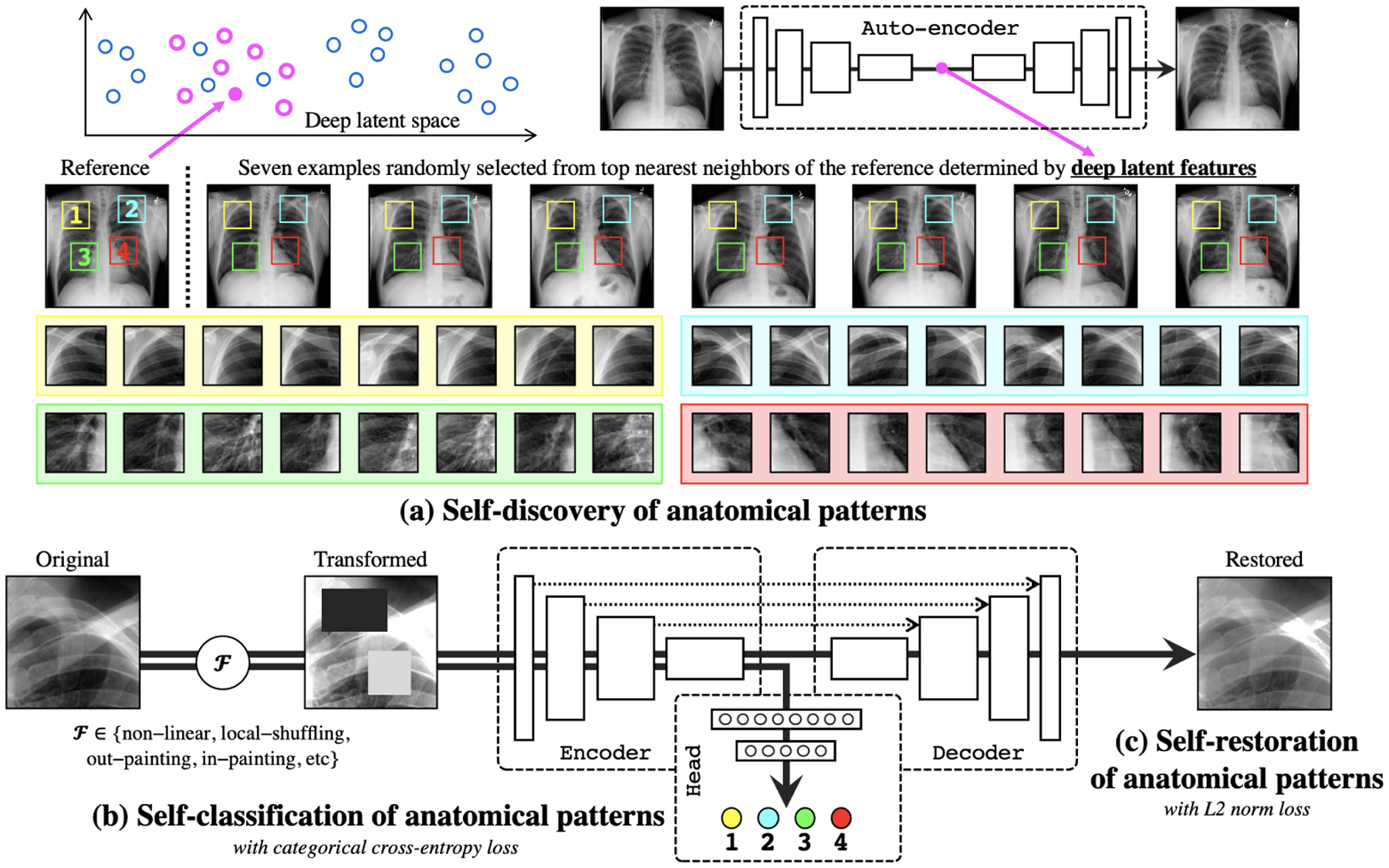
Medical images are naturally associated with rich semantics about the human anatomy, reflected in an abundance of recurring anatomical patterns, offering unique potential to foster deep semantic representation learning and yield semantically more powerful models for different medical applications. But how exactly such strong yet free semantics embedded in medical images can be harnessed for self-supervised learning remains largely unexplored. To this end, we train deep models to learn semantically enriched visual representation by self-discovery, self-classification, and self-restoration of the anatomy underneath medical images, resulting in a semantics-enriched, general-purpose, pre-trained 3D model, named Semantic Genesis. We examine our Semantic Genesis with all the publicly-available pre-trained models, by either self-supervision or fully supervision, on the six distinct target tasks, covering both classification and segmentation in various medical modalities (., CT, MRI, and X-ray). Our extensive experiments demonstrate that Semantic Genesis significantly exceeds all of its 3D counterparts as well as the ImageNet-based transfer learning in 2D. This performance is attributed to our novel self-supervised learning framework, encouraging deep models to learn compelling semantic representation from abundant anatomical patterns resulting from consistent anatomies embedded in medical images. Code and pre-trained Semantic Genesis are available at https://github.com/JLiangLab/SemanticGenesis.

摘要

医学图像自然地与有关人体解剖结构的丰富语义相关联,这体现在大量反复出现的解剖模式中,为促进深度语义表征学习以及为不同医学应用生成语义更强大的模型提供了独特潜力。但是,如何确切地利用医学图像中嵌入的这种强大而自由的语义进行自监督学习,在很大程度上仍未得到探索。为此,我们训练深度模型,通过对医学图像下方解剖结构的自我发现、自我分类和自我恢复来学习语义丰富的视觉表征,从而得到一个名为语义起源(Semantic Genesis)的语义丰富、通用的预训练3D模型。我们在六个不同的目标任务上,通过自监督或完全监督,将我们的语义起源模型与所有公开可用的预训练模型进行比较,这些任务涵盖了各种医学模态(如CT、MRI和X射线)中的分类和分割。我们广泛的实验表明,语义起源模型显著超越了所有其他3D对应模型以及基于ImageNet的2D迁移学习。这种性能归因于我们新颖的自监督学习框架,该框架鼓励深度模型从医学图像中嵌入的一致解剖结构所产生的丰富解剖模式中学习引人注目的语义表征。代码和预训练的语义起源模型可在https://github.com/JLiangLab/SemanticGenesis获取。

https://cdn.ncbi.nlm.nih.gov/pmc/blobs/e26c/9187496/75cfcd2d9e6a/nihms-1812896-f0003.jpg
https://cdn.ncbi.nlm.nih.gov/pmc/blobs/e26c/9187496/cb4ed1a44def/nihms-1812896-f0004.jpg
https://cdn.ncbi.nlm.nih.gov/pmc/blobs/e26c/9187496/5b07b2cb580e/nihms-1812896-f0005.jpg
https://cdn.ncbi.nlm.nih.gov/pmc/blobs/e26c/9187496/7471b42e4ef4/nihms-1812896-f0001.jpg
https://cdn.ncbi.nlm.nih.gov/pmc/blobs/e26c/9187496/e9f201657e01/nihms-1812896-f0002.jpg
https://cdn.ncbi.nlm.nih.gov/pmc/blobs/e26c/9187496/75cfcd2d9e6a/nihms-1812896-f0003.jpg