Department of Molecular Pathophysiology, Institute for Advanced Medical Sciences, Nippon Medical School, Tokyo, Japan.
Department of Cell Biology, National Cerebral and Cardiovascular Center Research Institute, Osaka, Japan.
Kidney360. 2022 Jan 19;3(4):700-713. doi: 10.34067/KID.0005962021. eCollection 2022 Apr 28.
The renal glomerulus is a tuft of capillaries in Bowman's capsule and functions as a blood-filtration unit in the kidney. The unique glomerular capillary tuft structure is relatively conserved through vertebrate species. However, the morphogenetic mechanism governing glomerular capillary tuft formation remains elusive.
To clarify how glomerular capillaries develop, we analyzed glomerular capillary formation in the zebrafish pronephros by exploiting fluorescence-based bio-imaging technology.
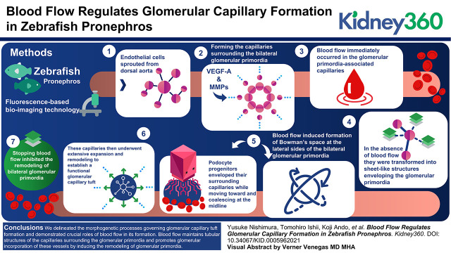
During glomerular capillary formation in the zebrafish pronephros, endothelial cells initially sprouted from the dorsal aorta and formed the capillaries surrounding the bilateral glomerular primordia in response to podocyte progenitor-derived vascular endothelial growth factor-A. After formation, blood flow immediately occurred in the glomerular primordia-associated capillaries, while in the absence of blood flow, they were transformed into sheet-like structures enveloping the glomerular primordia. Subsequently, blood flow induced formation of Bowman's space at the lateral sides of the bilateral glomerular primordia. Concomitantly, podocyte progenitors enveloped their surrounding capillaries while moving toward and coalescing at the midline. These capillaries then underwent extensive expansion and remodeling to establish a functional glomerular capillary tuft. However, stopping blood flow inhibited the remodeling of bilateral glomerular primordia, which therefore remained unvascularized but covered by the vascular sheets.
We delineated the morphogenetic processes governing glomerular capillary tuft formation in the zebrafish pronephros and demonstrated crucial roles of blood flow in its formation. Blood flow maintains tubular structures of the capillaries surrounding the glomerular primordia and promotes glomerular incorporation of these vessels by inducing the remodeling of glomerular primordia.
肾小球是鲍曼囊中的毛细血管丛,作为肾脏中的血液过滤单位。脊椎动物物种中相对保守的独特肾小球毛细血管丛结构。然而,控制肾小球毛细血管丛形成的形态发生机制仍然难以捉摸。
为了阐明肾小球毛细血管是如何发育的,我们利用基于荧光的生物成像技术分析了斑马鱼前肾中的肾小球毛细血管形成。
在斑马鱼前肾的肾小球毛细血管形成过程中,内皮细胞最初从背主动脉发芽,并响应足细胞祖细胞衍生的血管内皮生长因子-A 形成围绕双侧肾小球原基的毛细血管。形成后,血流立即发生在肾小球原基相关的毛细血管中,而在没有血流的情况下,它们转化为包裹肾小球原基的片状结构。随后,血流诱导在双侧肾小球原基的侧部形成鲍曼氏腔。同时,足细胞祖细胞在向中线移动并融合的过程中包裹其周围的毛细血管。这些毛细血管随后经历广泛的扩张和重塑,以建立功能性的肾小球毛细血管丛。然而,停止血流会抑制双侧肾小球原基的重塑,因此这些原基仍然没有血管化,但被血管片覆盖。
我们描绘了斑马鱼前肾中肾小球毛细血管丛形成的形态发生过程,并证明了血流在其形成中的关键作用。血流维持围绕肾小球原基的毛细血管的管状结构,并通过诱导肾小球原基的重塑来促进这些血管的肾小球整合。