Bagio Dini Asrianti, Julianto Indah, Margono Anggraini, Suprastiwi Endang
Department of Conservative Dentistry, Faculty of Dentistry, Universitas Indonesia, Jakarta, Indonesia.
Department of Dermato Venereology, Faculty of Medicine, Universitas Sebelas Maret, Solo Surakarta, Indonesia.
Eur J Dent. 2023 Feb;17(1):173-182. doi: 10.1055/s-0042-1744370. Epub 2022 Jun 21.
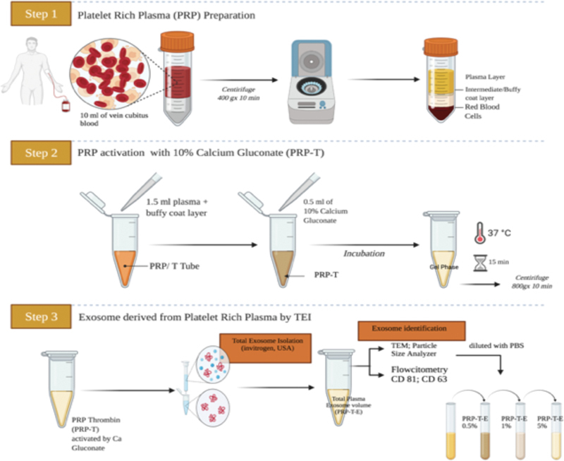
This study analyzed the potential of various concentrations of the thrombin-activated platelet-derived exosome (T-aPDE) to regenerate the dental pulp by performing an analysis of the cell viability, migration activity, and vascular endothelial growth factor A (VEGF-A) expression of human dental pulp stem cells (hDPSCs).
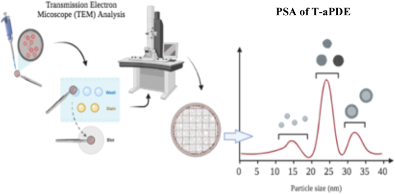
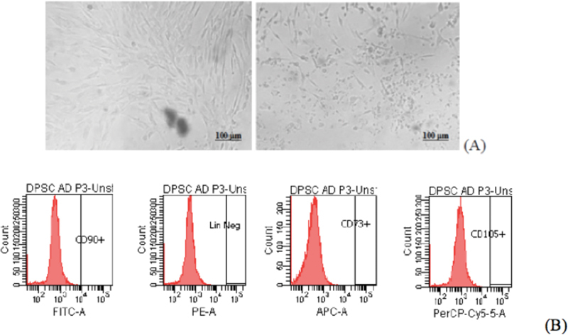
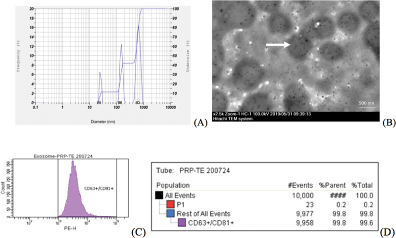
The hDPSCs were collected from nine third molar teeth of nine healthy donors and were isolated and cultured using the explant method. They were harvested between the third and fourth passages and starved, after which they were seeded in the following treatments: Dulbecco's Modified Eagle Medium and 10% platelet-rich plasma-thrombin as the control groups, and 0.5, 1, and 5% T-aPDE as the experimental groups. All groups had three biological triplicates (Triplo) and two number of experiments. The T-aPDE was analyzed using transmission electron microscopy testing, particle size analyzer, and CD63 + and CD81 + specific immune phenotyping flow cytometry tests for plasma exosomes. The cell viability was evaluated using the colorimetric assay of activity cellular enzymes (MTT assay); the migration activity, using scratch assay; and the VEGF-A expression, using enzyme-linked immunosorbent assay.
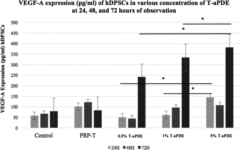
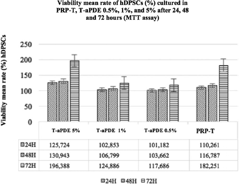
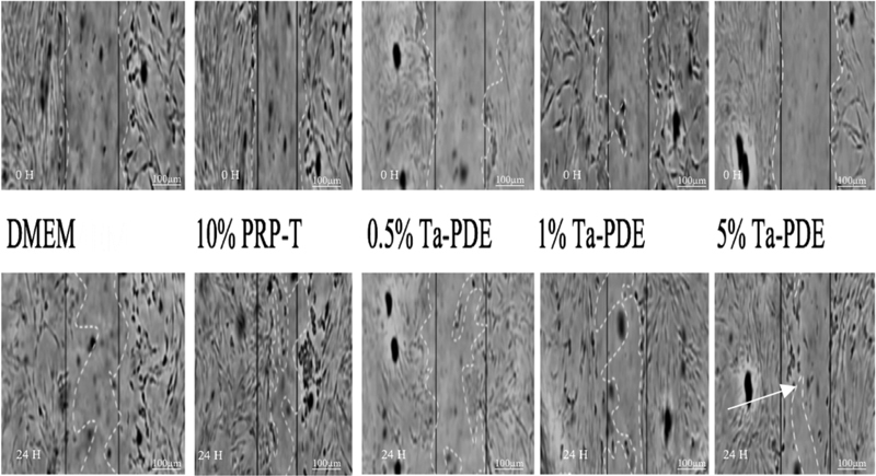
The highest viability absorbance value of hDPSCs after 24, 48, 72 hours of observation was in the 5% T-aPDE group (<0.05). Whereas, the closest distance result of migratory activation hDPSCs was also in the same group (<0.05). However the highest VEGF-A expression of hDSPCs was noted in the same group at 72 hours observation (<0.05).
The data were analyzed using one-way analysis of variance and the Kruskal-Wallis test. The statistical power was set at <0.05 CONCLUSION: The 5% T-aPDE had a higher potential to induce dental pulp regeneration than the other groups.
本研究通过分析人牙髓干细胞(hDPSC)的细胞活力、迁移活性和血管内皮生长因子A(VEGF-A)表达,来分析不同浓度的凝血酶激活的血小板衍生外泌体(T-aPDE)对牙髓再生的潜力。
从9名健康供体的9颗第三磨牙中收集hDPSC,采用组织块法进行分离培养。在第3代和第4代之间收获细胞并进行饥饿处理,之后将其接种于以下处理中:杜氏改良 Eagle 培养基和10%富血小板血浆-凝血酶作为对照组,0.5%、1%和5% T-aPDE作为实验组。所有组均有3个生物学重复(Triplo)和2次实验。使用透射电子显微镜检测、粒度分析仪以及针对血浆外泌体的CD63 +和CD81 +特异性免疫表型流式细胞术检测来分析T-aPDE。使用细胞活性酶比色测定法(MTT法)评估细胞活力;使用划痕试验评估迁移活性;使用酶联免疫吸附测定法评估VEGF-A表达。
观察24、48、72小时后,hDPSC的最高活力吸光度值出现在5% T-aPDE组(<0.05)。而hDPSC迁移激活的最接近距离结果也在同一组(<0.05)。然而,在观察72小时时,hDSPCs的最高VEGF-A表达出现在同一组(<0.05)。
使用单因素方差分析和Kruskal-Wallis检验对数据进行分析。设定统计功效<0.05。结论:5% T-aPDE比其他组具有更高的诱导牙髓再生的潜力。