Department of Applied Physics, Experimental Biomolecular Physics, Albanova Univ Center, Royal Institute of Technology (KTH), 106 91, Stockholm, Sweden.
Department of Oncology-Pathology, K7, Z1:00, Karolinska University Hospital, Karolinska Institutet, 171 76, Stockholm, Sweden.
J Nanobiotechnology. 2022 Jun 21;20(1):292. doi: 10.1186/s12951-022-01502-w.
Increasing evidence suggests that platelets play a central role in cancer progression, with altered storage and selective release from platelets of specific tumor-promoting proteins as a major mechanism. Fluorescence-based super-resolution microscopy (SRM) can resolve nanoscale spatial distribution patterns of such proteins, and how they are altered in platelets upon different activations. Analysing such alterations by SRM thus represents a promising, minimally invasive strategy for platelet-based diagnosis and monitoring of cancer progression. However, broader applicability beyond specialized research labs will require objective, more automated imaging procedures. Moreover, for statistically significant analyses many SRM platelet images are needed, of several different platelet proteins. Such proteins, showing alterations in their distributions upon cancer progression additionally need to be identified.
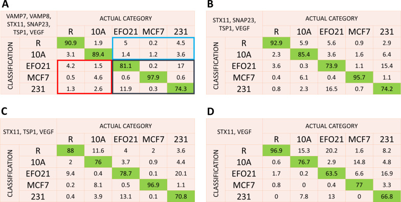
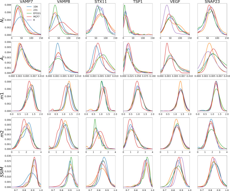
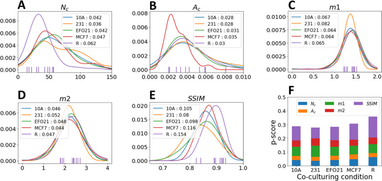
A fast, streamlined and objective procedure for SRM platelet image acquisition, analysis and classification was developed to overcome these limitations. By stimulated emission depletion SRM we imaged nanoscale patterns of six different platelet proteins; four different SNAREs (soluble N-ethylmaleimide factor attachment protein receptors) mediating protein secretion by membrane fusion of storage granules, and two angiogenesis regulating proteins, representing cargo proteins within these granules coupled to tumor progression. By a streamlined procedure, we recorded about 100 SRM images of platelets, for each of these six proteins, and for five different categories of platelets; incubated with cancer cells (MCF-7, MDA-MB-231, EFO-21), non-cancer cells (MCF-10A), or no cells at all. From these images, structural similarity and protein cluster parameters were determined, and probability functions of these parameters were generated for the different platelet categories. By comparing these probability functions between the categories, we could identify nanoscale alterations in the protein distributions, allowing us to classify the platelets into their correct categories, if they were co-incubated with cancer cells, non-cancer cells, or no cells at all.
The fast, streamlined and objective acquisition and analysis procedure established in this work confirms the role of SNAREs and angiogenesis-regulating proteins in platelet-mediated cancer progression, provides additional fundamental knowledge on the interplay between tumor cells and platelets, and represent an important step towards using tumor-platelet interactions and redistribution of nanoscale protein patterns in platelets as a basis for cancer diagnostics.
越来越多的证据表明,血小板在癌症进展中起着核心作用,其改变的储存和从血小板中选择性释放特定的促进肿瘤生长的蛋白质是主要机制。基于荧光的超分辨率显微镜(SRM)可以解析这些蛋白质的纳米级空间分布模式,以及它们在不同激活条件下在血小板中的改变。因此,通过 SRM 分析这些改变代表了一种有前途的、微创的基于血小板的癌症进展诊断和监测策略。然而,要在更广泛的范围内应用,除了专门的研究实验室外,还需要客观的、更自动化的成像程序。此外,为了进行统计学上有意义的分析,需要对几种不同的血小板蛋白进行大量的 SRM 血小板图像分析。这些蛋白质在癌症进展过程中其分布发生改变,还需要对其进行鉴定。
为了克服这些限制,我们开发了一种快速、简化和客观的 SRM 血小板图像采集、分析和分类的方法。通过受激发射损耗 SRM,我们对六种不同的血小板蛋白的纳米级模式进行成像;其中四种 SNARE(可溶性 N-乙基马来酰亚胺因子附着蛋白受体)通过膜融合介导储存颗粒中的蛋白质分泌,两种血管生成调节蛋白,代表这些颗粒内与肿瘤进展相关的货物蛋白。通过简化的程序,我们记录了大约 100 张血小板的 SRM 图像,每张图像代表这六种蛋白质中的一种,以及五种不同类别的血小板;与癌细胞(MCF-7、MDA-MB-231、EFO-21)、非癌细胞(MCF-10A)或根本没有细胞孵育。从这些图像中,确定了结构相似性和蛋白质簇参数,并为不同的血小板类别生成了这些参数的概率函数。通过比较这些类别之间的概率函数,我们可以识别蛋白质分布的纳米级改变,从而将血小板正确分类为它们与癌细胞、非癌细胞或根本没有细胞孵育的类别。
本工作中建立的快速、简化和客观的获取和分析程序证实了 SNARE 和血管生成调节蛋白在血小板介导的癌症进展中的作用,为肿瘤细胞与血小板之间的相互作用以及肿瘤-血小板相互作用和血小板中纳米级蛋白质模式的重新分布提供了更多的基础知识,作为癌症诊断的基础。