Institute of Biochemistry and Molecular Medicine, Faculty of Medicine, University of Bern, 3012 Bern, Switzerland.
Swiss National Center of Competence in Research, NCCR TransCure, University of Bern, 3012 Bern, Switzerland.
Cells. 2022 Jun 11;11(12):1898. doi: 10.3390/cells11121898.
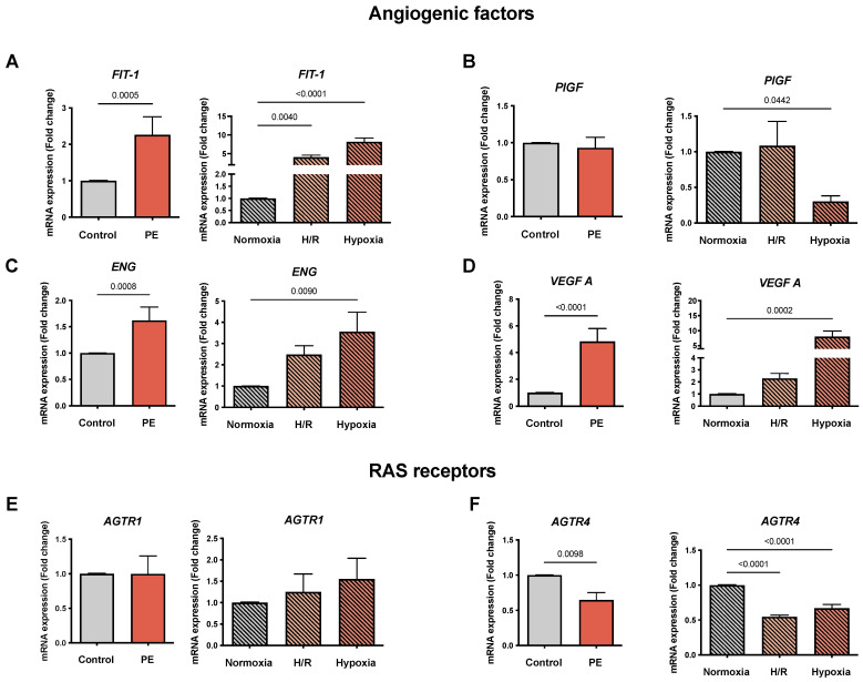
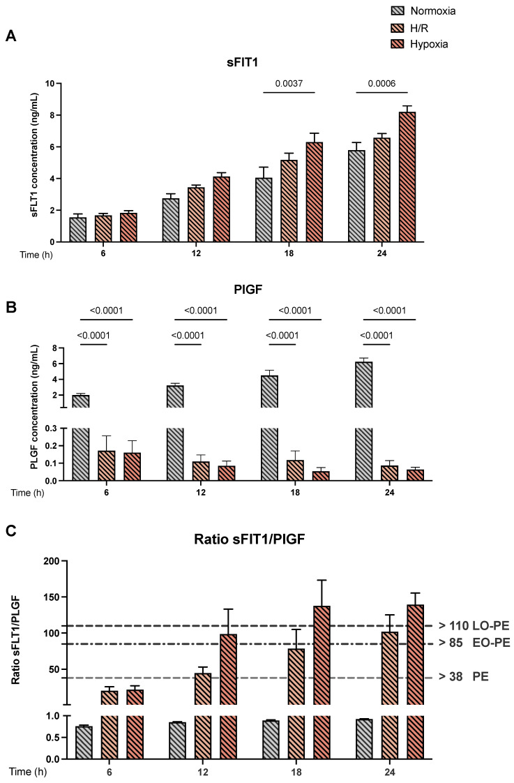
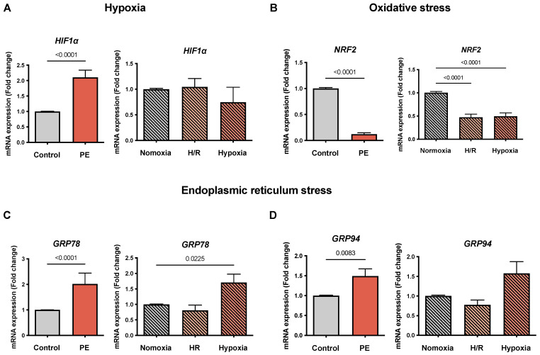
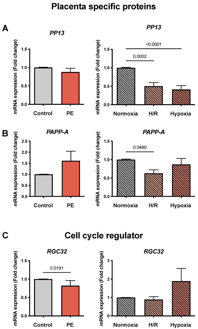
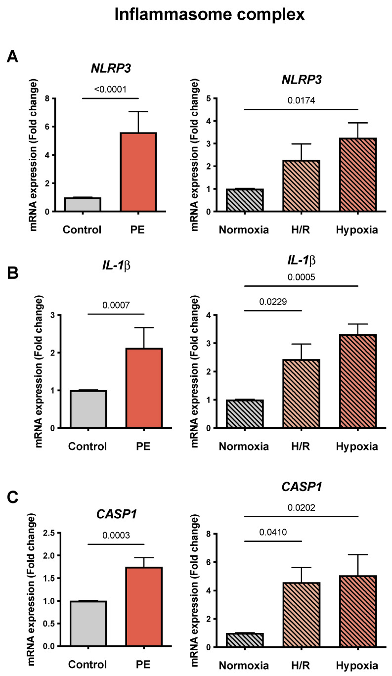
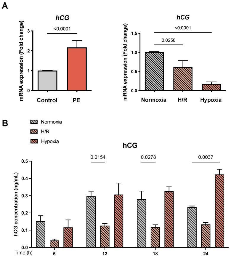
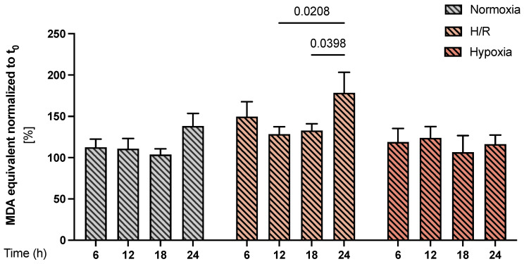
Preeclampsia (PE) is a pregnancy-specific disorder that affects 3 to 5% of pregnancies worldwide and is one of the leading causes of maternal and fetal morbidity and mortality. Nevertheless, how these events occur remains unclear. We hypothesized that the induction of hypoxic conditions in vitro in primary human trophoblast cells would mimic several characteristics of PE found in vivo. We applied and characterized a model of primary cytotrophoblasts isolated from healthy pregnancies that were placed under different oxygen concentrations: ambient O (5% pCO, 21%pO, 24 h, termed "normoxia"), low O concentration (5% pCO, 1.5% pO, 24 h, termed "hypoxia"), or "hypoxia/reoxygenation" (H/R: 6 h intervals of normoxia and hypoxia for 24 h). Various established preeclamptic markers were assessed in this cell model and compared to placental tissues obtained from PE pregnancies. Seventeen PE markers were analyzed by qPCR, and the protein secretion of soluble fms-like tyrosine kinase 1 (sFlT-1) and the placenta growth factor (PlGF) was determined by ELISA. Thirteen of seventeen genes associated with angiogenesis, the renin-angiotensin system, oxidative stress, endoplasmic reticulum stress, and the inflammasome complex were susceptible to H/R and hypoxia, mimicking the expression pattern of PE tissue. In cell culture supernatants, the secretion of sFlT-1 was increased in hypoxia, while PlGF release was significantly reduced in H/R and hypoxia. In the supernatants of our cell models, the sFlT-1/PlGF ratio in hypoxia and H/R was higher than 38, which is a strong indicator for PE in clinical practice. These results suggest that our cellular models reflect important pathological processes occurring in PE and are therefore suitable as PE in vitro models.
子痫前期 (PE) 是一种妊娠特有的疾病,影响全球 3%至 5%的妊娠,是孕产妇和胎儿发病率和死亡率的主要原因之一。然而,这些事件的发生机制仍不清楚。我们假设,在体外原代人滋养细胞中诱导缺氧条件将模拟体内发现的几种 PE 特征。我们应用并描述了一种从健康妊娠中分离的原代滋养细胞的模型,这些细胞置于不同的氧浓度下:大气氧(5% pCO,21% pO,24 小时,称为“常氧”)、低氧浓度(5% pCO,1.5% pO,24 小时,称为“缺氧”)或“缺氧/复氧”(H/R:24 小时内进行 6 小时的常氧和缺氧间隔)。在这个细胞模型中评估了各种已建立的子痫前期标志物,并与从 PE 妊娠中获得的胎盘组织进行了比较。通过 qPCR 分析了 17 个 PE 标志物,通过 ELISA 测定了可溶性 fms 样酪氨酸激酶 1 (sFlT-1) 和胎盘生长因子 (PlGF) 的蛋白分泌。与血管生成、肾素-血管紧张素系统、氧化应激、内质网应激和炎症小体复合物相关的 13 个基因易受 H/R 和缺氧的影响,模拟了 PE 组织的表达模式。在细胞培养上清液中,缺氧时 sFlT-1 的分泌增加,而 H/R 和缺氧时 PlGF 的释放显著减少。在我们的细胞模型上清液中,缺氧和 H/R 中的 sFlT-1/PlGF 比值高于 38,这是临床实践中 PE 的一个强烈指标。这些结果表明,我们的细胞模型反映了 PE 中发生的重要病理过程,因此适合作为 PE 的体外模型。