Department of Infectious Diseases and Hepatology, Nanfang Hospital, Southern Medical University, Guangzhou, China.
Department of Oncology, Nanfang Hospital, Southern Medical University, Guangzhou, China.
Front Immunol. 2022 Jun 9;13:868809. doi: 10.3389/fimmu.2022.868809. eCollection 2022.
The clinical significance of liver stiffness (LS) measured by shear wave elastography (SWE) in programmed cell death protein-1 (PD-1) inhibitors treated advanced hepatocellular carcinoma (HCC) patients remains unknown. This study aimed to explore the prognostic value of baseline LS by SWE prior to PD-1 inhibitor treatment in combination with lenvatinib.
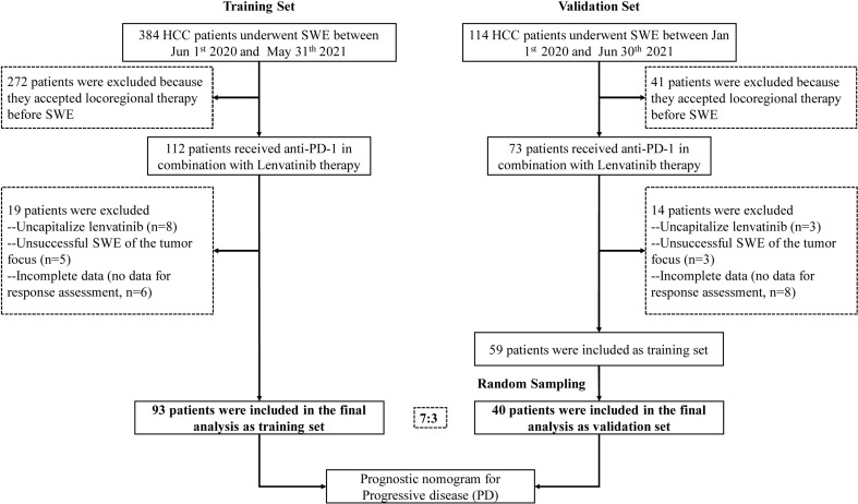
We retrospectively evaluated patients (n=133) with HCC who received anti-PD-1 antibodies plus lenvatinib at two high-volume medical centres, between January 2020 and June 2021. Univariate and multivariate logistic regression analysis were used to develop a novel nomogram. RNA sequencing and immunohistochemical staining were used to assess the heterogeneity of biological and immune characteristics associated with tumor stiffness.
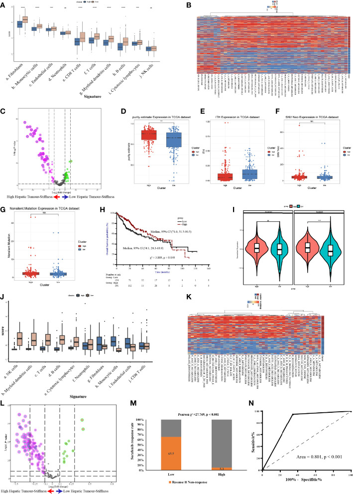
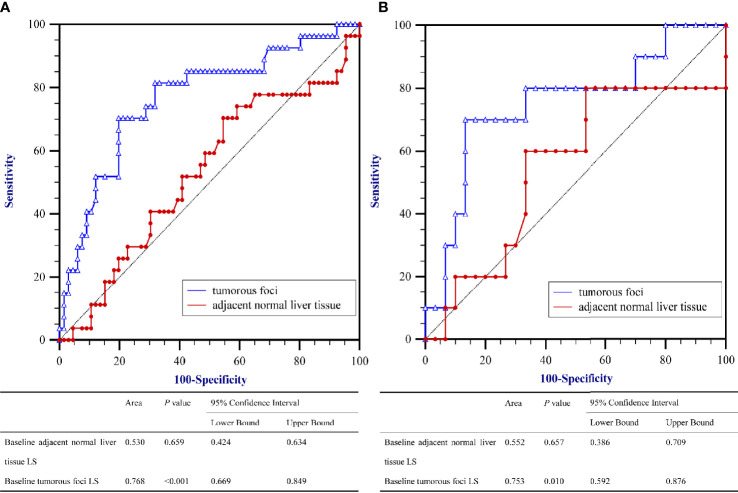
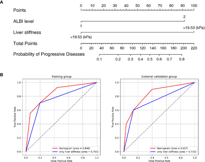
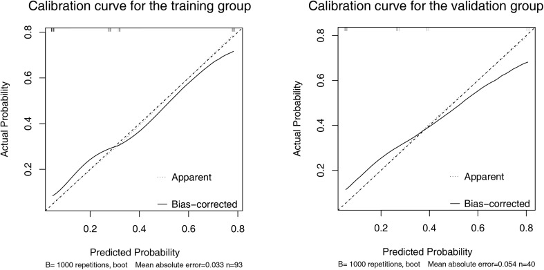
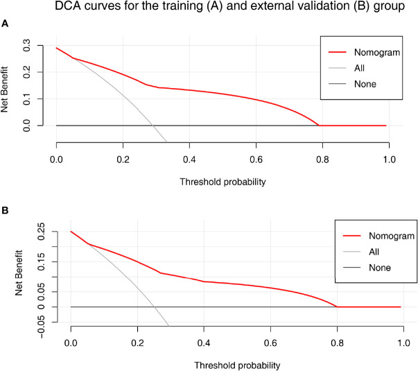
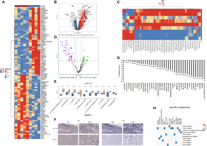
The objective response rate (ORR) and disease control rate (DCR) of the whole population were 23.4% and 72.2%, respectively. A LS value of the baseline tumorous foci of 19.53 kPa had the maximum sum of sensitivity and specificity, making it the optimal cut-off value for predicting PD-1 inhibitor efficacy. The nomogram comprised baseline tumor LS and albumin-bilirubin grade (ALBI), which provided favorable calibration and discrimination in the training dataset with an AUC of 0.840 (95%CI: 0.750-0.931) and a C-index of 0.828. Further, it showed acceptable discrimination in the validation cohort, with an AUC of 0.827 (95%CI: 0.673-0.980) and C-index of 0.803. The differentially expressed genes enriched in high stiffness tumors were predominantly associated with metabolic pathways, while those enriched in low stiffness tumors were related to DNA damage repair. Furthermore, patients with high stiffness tumors had a relatively lower infiltration of immune cells and histone deacetylase pathway inhibitors were identified as candidate drugs to promote the efficacy of immunotherapy.
Baseline LS value of tumorous foci by SWE-that is, before administration of a PD-1 inhibitor in combination with lenvatinib-is a convenient predictor of PD-1 inhibitor efficacy in patients with advanced HCC, which has potential to be used for pretreatment stratification to optimize treatment of advanced HCC.
程序性细胞死亡蛋白-1(PD-1)抑制剂治疗晚期肝细胞癌(HCC)患者时,通过剪切波弹性成像(SWE)测量的肝硬度(LS)的临床意义尚不清楚。本研究旨在探讨 PD-1 抑制剂联合仑伐替尼治疗前 SWE 基线 LS 在预测疗效方面的价值。
我们回顾性评估了 2020 年 1 月至 2021 年 6 月期间在两家高水平医疗中心接受抗 PD-1 抗体联合仑伐替尼治疗的 HCC 患者(n=133)。采用单因素和多因素逻辑回归分析建立新的列线图。RNA 测序和免疫组织化学染色用于评估与肿瘤硬度相关的生物学和免疫特征的异质性。
全人群的客观缓解率(ORR)和疾病控制率(DCR)分别为 23.4%和 72.2%。基线肿瘤病灶 LS 值为 19.53 kPa 时,具有最大的敏感性和特异性之和,是预测 PD-1 抑制剂疗效的最佳截断值。列线图包含基线肿瘤 LS 和白蛋白-胆红素分级(ALBI),在训练数据集中具有良好的校准度和区分度,AUC 为 0.840(95%CI:0.750-0.931),C 指数为 0.828。此外,在验证队列中也具有可接受的区分度,AUC 为 0.827(95%CI:0.673-0.980),C 指数为 0.803。高硬度肿瘤中富集的差异表达基因主要与代谢途径相关,而低硬度肿瘤中富集的基因与 DNA 损伤修复相关。此外,高硬度肿瘤患者的免疫细胞浸润程度相对较低,组蛋白去乙酰化酶抑制剂被鉴定为促进免疫治疗疗效的候选药物。
SWE 测量的肿瘤病灶基线 LS 值(即 PD-1 抑制剂联合仑伐替尼治疗前)是晚期 HCC 患者 PD-1 抑制剂疗效的一个方便的预测指标,具有用于治疗前分层以优化晚期 HCC 治疗的潜力。