Federal Technological University of Paraná/Graduate Program in Electrical Engineering and Industrial Informatics, Endereço: Av. Sete de Setembro 3165. Campus Curitiba Sede Centro, Curitiba, PR, CEP-80230-901, Brazil.
Federal Technological University of Paraná/Program in Biomedical Engineering, Endereço: Av. Silva Jardim, 807 - Bloco V3. Campus Curitiba Sede Centro, Curitiba, PR, CEP-80230-901, Brasil.
Biomed Eng Online. 2022 Jun 27;21(1):41. doi: 10.1186/s12938-022-01009-3.
Considering the estimate that thyroid cancer will become the fourth most prevalent type of tumor, improving its diagnosis is a necessity. The gold standard for evaluating thyroid nodules is ultrasound followed by biopsy. These tests, however, have limitations, especially in nodules smaller than 0.5 cm. Dynamic infrared thermography is an imaging method that does not require ionizing radiation or contrast injection. The aim of the study was to analyze the thermal behavior of thyroid nodules through infrared thermography using the cold stress protocol.
The Wilcoxon test showed thermal differences between groups (control and healthy, p < 0.001). The difference in the thermal behavior of the nodular tissues was evidenced by the longitudinal analysis. When comparing the nodules, it was possible to verify that the beginnings of tissue heating is significant (p = 0.001). In addition, the variability analysis showed a "well" effect, which occurred in period t-1 (pre-cooling time) to period t = 3 (time three minutes). Benign nodules had a variation ratio of 1.81 compared to malignant nodules.
Benign nodules present a different thermal behavior than malignant nodules, and both present different behavior than normal tissue. For the analysis of nodules, the protocol used with cold stress, dynamic thermography and the inclusion of time t-1 were essential for the differentiation of nodules in the thyroid gland. Therefore, we recommend the continuance of these parameters for future studies.
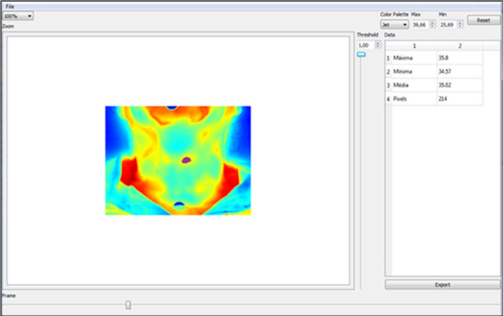
Thirty-three individuals with nodules in the thyroid region and nine healthy individuals participated in this descriptive exploratory study. In total, 42 nodules were evaluated, 11 malignant and 31 benign. The region of interest was exposed to cold stress for 30 s. First, the image was captured before the cold stress and subsequently, the images were assessed every 30 s, over a 10-min time period after cold stress. The perfusion and the thermal behavior of the tissues were evaluated by longitudinal analysis based on the number of pixels in each time period. The statistical tests of Wilcoxon, F-Snedecor and longitudinal models would assist in data analysis.
考虑到甲状腺癌将成为第四大常见肿瘤,提高其诊断水平是必要的。评估甲状腺结节的金标准是超声检查加活检。然而,这些检查存在局限性,特别是对于小于 0.5 厘米的结节。动态红外热像图是一种不需要电离辐射或造影剂注射的成像方法。本研究的目的是通过冷应激方案分析甲状腺结节的热行为。
Wilcoxon 检验显示,对照组和健康组之间存在热差异(p<0.001)。通过纵向分析证实了结节组织热行为的差异。在比较结节时,我们可以发现组织加热的开始具有显著差异(p=0.001)。此外,变异性分析显示出“良好”的效果,这种效果发生在 t-1 期(预冷却时间)到 t=3 期(三分钟时间)。良性结节的变异比为 1.81,而恶性结节为 1.71。
良性结节的热行为与恶性结节不同,而这两者与正常组织的热行为也不同。对于结节的分析,冷应激、动态热像图和包含 t-1 期的方案对于甲状腺结节的区分至关重要。因此,我们建议在未来的研究中继续使用这些参数。
本描述性探索性研究纳入了 33 名甲状腺区域有结节的个体和 9 名健康个体。共评估了 42 个结节,其中 11 个为恶性,31 个为良性。感兴趣区域暴露于冷应激 30 秒。首先,在冷应激前拍摄图像,然后在冷应激后每 30 秒拍摄一次图像,共拍摄 10 分钟。通过基于每个时间段的像素数的纵向分析来评估组织的灌注和热行为。Wilcoxon、F-Snedecor 和纵向模型的统计检验将有助于数据分析。