Department of Ophthalmology and Optometry, Medical University of Vienna, Vienna, Austria.
Eye (Lond). 2023 May;37(7):1439-1444. doi: 10.1038/s41433-022-02156-6. Epub 2022 Jul 1.
BACKGROUND/OBJECTIVES: We aim to develop an objective fully automated Artificial intelligence (AI) algorithm for MNV lesion size and leakage area segmentation on fluorescein angiography (FA) in patients with neovascular age-related macular degeneration (nAMD).
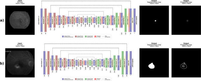
SUBJECTS/METHODS: Two FA image datasets collected form large prospective multicentre trials consisting of 4710 images from 513 patients and 4558 images from 514 patients were used to develop and evaluate a deep learning-based algorithm to detect CNV lesion size and leakage area automatically. Manual segmentation of was performed by certified FA graders of the Vienna Reading Center. Precision, Recall and F1 score between AI predictions and manual annotations were computed. In addition, two masked retina experts conducted a clinical-applicability evaluation, comparing the quality of AI based and manual segmentations.
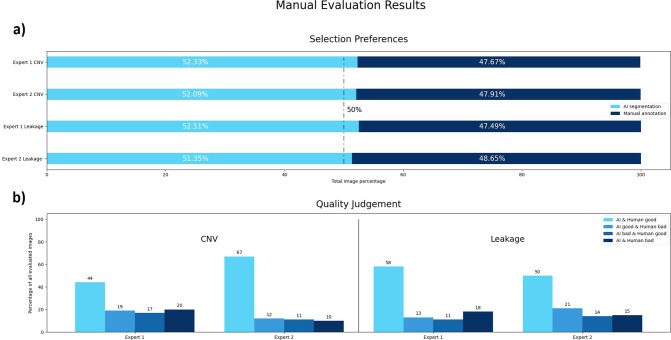
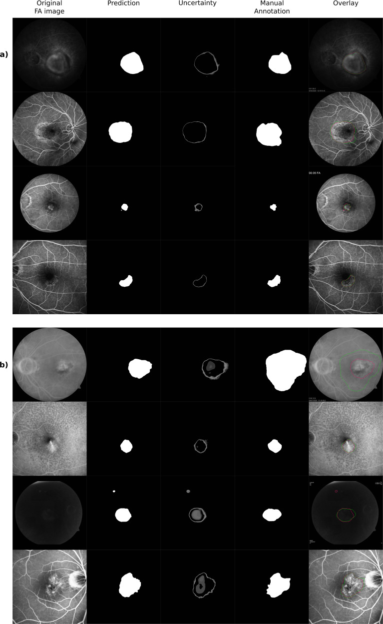
For CNV lesion size and leakage area segmentation, we obtained F1 scores of 0.73 and 0.65, respectively. Expert review resulted in a slight preference for the automated segmentations in both datasets. The quality of automated segmentations was slightly more often judged as good compared to manual annotations.
CNV lesion size and leakage area can be segmented by our automated model at human-level performance, its output being well-accepted during clinical applicability testing. The results provide proof-of-concept that an automated deep learning approach can improve efficacy of objective biomarker analysis in FA images and will be well-suited for clinical application.
背景/目的:我们旨在开发一种客观的全自动人工智能(AI)算法,用于对年龄相关性黄斑变性(AMD)新生血管患者的荧光素血管造影(FA)中的 MNV 病变大小和渗漏区进行分割。
受试者/方法:使用来自大型前瞻性多中心试验的两个 FA 图像数据集,包括 513 名患者的 4710 张图像和 514 名患者的 4558 张图像,用于开发和评估一种基于深度学习的算法,以自动检测 CNV 病变大小和渗漏区。由维也纳阅读中心的认证 FA 分级员进行手动分割。计算 AI 预测与手动注释之间的精度、召回率和 F1 分数。此外,两名受屏蔽的视网膜专家进行了临床适用性评估,比较了基于 AI 和手动分割的质量。
对于 CNV 病变大小和渗漏区分割,我们分别获得了 0.73 和 0.65 的 F1 分数。专家审查导致在两个数据集的自动分割中都有轻微偏好。与手动注释相比,自动分割的质量更经常被判断为良好。
我们的自动模型可以达到人类水平的性能来分割 CNV 病变大小和渗漏区,其输出在临床适用性测试中得到了很好的接受。结果提供了一个概念证明,即自动深度学习方法可以提高 FA 图像中客观生物标志物分析的效果,并且非常适合临床应用。