Satoh Yoko, Ishida Jiro, Inui Yoshitaka, Takenaka Akinori, Bando Shuji, Ishida Sayuri, Toyama Hiroshi
Imaging Center, Fujita Medical Innovation Center Tokyo, Ota-ku 144-0041, Tokyo, Japan.
Department of Radiology, Faculty of Medicine, Fujita Health University, Toyoake 470-1192, Aichi, Japan.
Diagnostics (Basel). 2024 Sep 19;14(18):2068. doi: 10.3390/diagnostics14182068.
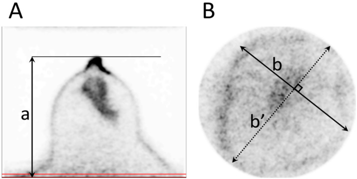
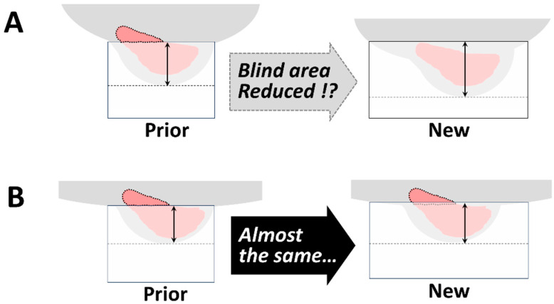
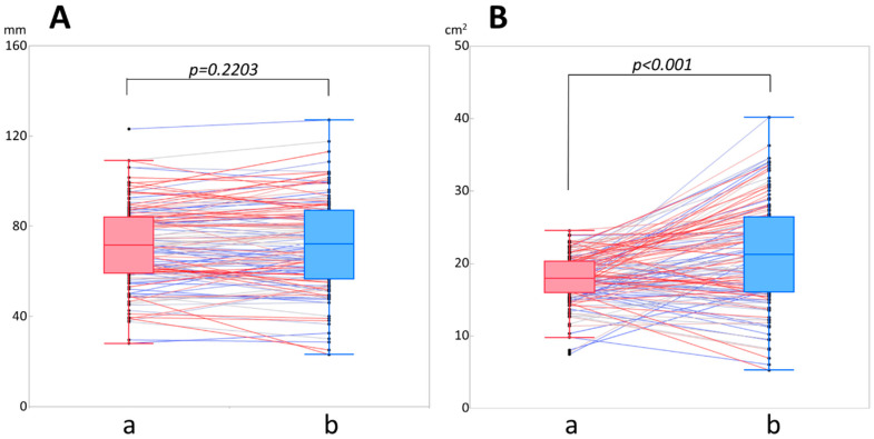
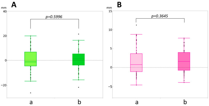
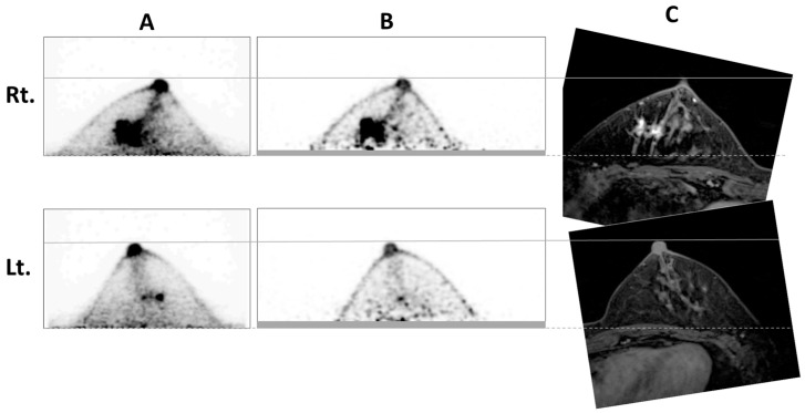
: Breast-specific positron emission tomography (PET) provides higher sensitivity and spatial resolution than whole-body PET/CT, but it has a blind area. Mammary glands near the chest wall sometimes present outside the field of view (FOV). A newer, dedicated breast PET (dbPET) model has a cylindrical detector with a larger diameter than previous models, so it is expected to eliminate or reduce blind areas. This study aimed to compare breast images acquired on the new dbPET model with images acquired on an older dbPET model to evaluate blind area reduction. : The nipple-to-chest wall distance (mm), maximum breast cross-sectional area at the FOV edge (cm) on the dbPET transverse images of the scanners, and the effects of patient age and body mass index (BMI) were compared. : There was no significant difference in the nipple-to-chest wall distance between the models ( = 0.223). The maximum breast cross-sectional area at the FOV edge was significantly larger on the newer model's images ( < 0.001). There was no significant correlation between breast size and the rate of change in both parameters. : The new ring-type dbPET scanners with larger diameter detectors did not reduce the blind area observed on older dbPET scanners.
乳腺专用正电子发射断层扫描(PET)比全身PET/CT具有更高的灵敏度和空间分辨率,但它存在一个盲区。靠近胸壁的乳腺有时会出现在视野(FOV)之外。一种更新的专用乳腺PET(dbPET)模型具有比以前型号直径更大的圆柱形探测器,因此有望消除或减少盲区。本研究旨在比较在新的dbPET模型上获取的乳腺图像与在旧的dbPET模型上获取的图像,以评估盲区的减少情况。:比较了乳头到胸壁的距离(mm)、扫描仪dbPET横向图像上FOV边缘处的最大乳腺横截面积(cm)以及患者年龄和体重指数(BMI)的影响。:两种模型之间乳头到胸壁的距离没有显著差异( = 0.223)。新模型图像上FOV边缘处的最大乳腺横截面积明显更大( < 0.001)。乳腺大小与两个参数的变化率之间没有显著相关性。:具有更大直径探测器的新型环形dbPET扫描仪并未减少在旧的dbPET扫描仪上观察到的盲区。