Moredun Research Institute, Pentlands Science Park, Penicuik, United Kingdom.
Roslin Institute, Royal (Dick) School of Veterinary Studies, University of Edinburgh, Penicuik, United Kingdom.
Front Cell Infect Microbiol. 2022 Jun 30;12:904606. doi: 10.3389/fcimb.2022.904606. eCollection 2022.
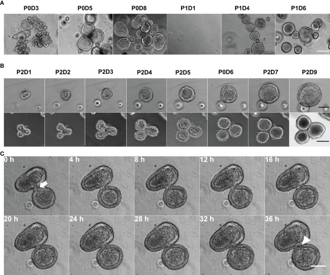
Gastro-intestinal nematode (GIN) parasites are a major cause of production losses in grazing cattle, primarily through reduced growth rates in young animals. Control of these parasites relies heavily on anthelmintic drugs; however, with growing reports of resistance to currently available anthelmintics, alternative methods of control are required. A major hurdle in this work has been the lack of physiologically relevant infection models that has made studying precise interactions between the host and the GINs difficult. Such mechanistic insights into the infection process will be valuable for the development of novel targets for drugs, vaccines, or other interventions. Here we created bovine gastric epithelial organoids from abomasal gastric tissue and studied their application as models for understanding host invasion by GIN parasites. Transcriptomic analysis of gastric organoids across multiple passages and the corresponding abomasal tissue showed conserved expression of tissue-specific genes across samples, demonstrating that the organoids are representative of bovine gastric tissue from which they were derived. We also show that self-renewing and self-organising three-dimensional organoids can also be serially passaged, cryopreserved, and resuscitated. Using , the most pathogenic gastric parasite in cattle in temperate regions, we show that cattle gastric organoids are biologically relevant models for studying GIN invasion in the bovine abomasum. Within 24 h of exposure, exsheathed larvae rapidly and repeatedly infiltrated the lumen of the organoids. Prior to invasion by the parasites, the abomasal organoids rapidly expanded, developing a 'ballooning' phenotype. Ballooning of the organoids could also be induced in response to exposure to parasite excretory/secretory products. In summary, we demonstrate the power of using abomasal organoids as a physiologically relevant model system to study interactions of and other GIN with bovine gastrointestinal epithelium.
胃肠线虫(GIN)寄生虫是放牧牛生产损失的主要原因,主要是通过降低幼畜的生长速度。这些寄生虫的控制主要依赖于驱虫药物;然而,随着越来越多的关于现有驱虫药物耐药性的报告,需要替代的控制方法。这项工作的一个主要障碍是缺乏与生理相关的感染模型,这使得研究宿主与 GIN 之间的精确相互作用变得困难。对感染过程的这种机制性了解对于开发新的药物、疫苗或其他干预措施的靶点将是有价值的。在这里,我们从皱胃胃组织中创建了牛胃上皮类器官,并研究了它们作为理解 GIN 寄生虫宿主入侵的模型的应用。在多个传代过程中对胃类器官进行转录组分析,并与相应的皱胃组织进行比较,结果表明组织特异性基因在样本中具有保守表达,这表明类器官代表了它们所来源于的牛胃组织。我们还表明,自我更新和自我组织的三维类器官也可以进行连续传代、冷冻保存和复苏。使用,在温带地区最具致病性的牛胃寄生虫,我们表明牛胃类器官是研究牛皱胃中 GIN 入侵的生物学相关模型。在暴露后 24 小时内,脱壳幼虫迅速而反复地渗透到类器官的腔中。在寄生虫入侵之前,皱胃类器官迅速扩张,形成“气球样”表型。暴露于寄生虫排泄/分泌产物也可以诱导类器官的气球样扩张。总之,我们证明了使用皱胃类器官作为生理相关模型系统来研究和其他 GIN 与牛胃肠道上皮相互作用的强大功能。