Dave Amisha D, Thavikulwat Alisa T, De Silva Tharindu, Wiley Henry E, Keenan Tiarnan D L, Wong Wai T, Cukras Catherine A
National Eye Institute, National Institutes of Health, Bethesda, MD, 20892, USA.
Am J Ophthalmol Case Rep. 2022 Jul 1;27:101647. doi: 10.1016/j.ajoc.2022.101647. eCollection 2022 Sep.
To perform longitudinal analysis of retinal arterial macroaneurysms in 3 patients with adult-onset Coats disease.
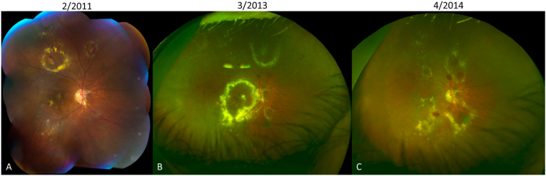
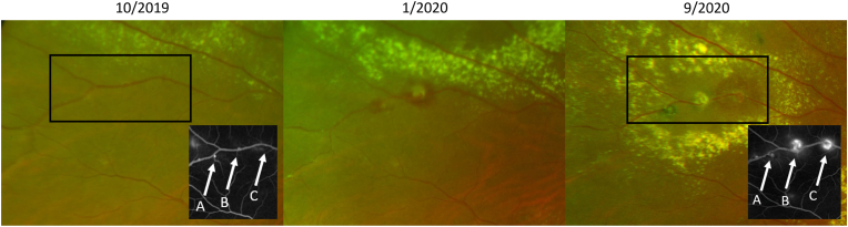
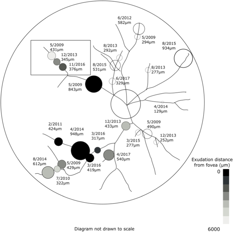
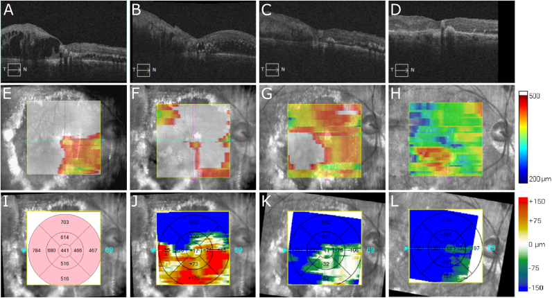
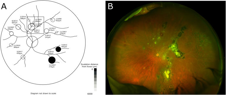
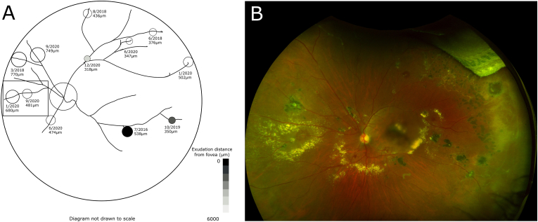
Three eyes of three patients with adult-onset Coats disease were followed longitudinally for 4-15 years. Ultra-widefield images and montage color fundus photographs of affected eyes were analyzed. Size, retinal location, and grading for predominant characteristic (hemorrhagic, exudative, or quiescent) of each individual macroaneurysm were followed longitudinally from the time of onset. Fifty-one individual retinal arterial macroaneurysms were identified. The distance of any lesion-associated hemorrhage or exudation present from the foveal center was measured. Macroaneurysms were located in all quadrants of the retina, with the majority (37/51) graded as hemorrhagic at lesion onset. Hemorrhagic and exudative macroaneurysms that entered the quiescent phase remained quiescent for an average of 26 months. Seven macroaneurysms were found to have hemorrhage or exudation that came within 125 μm of the fovea and all three eyes followed demonstrated a longitudinal decrease in visual acuity despite laser and intravitreal injection therapy. At the initial visit, visual acuities ranged from 20/40 to 20/200, but decreased to 20/80 to 20/320 by the last follow-up visit.
There are many challenges in treating patients with adult-onset Coats disease. Long-term loss of visual acuity often results from sequelae of hemorrhage and exudation affecting the macula.
对3例成人型Coats病患者的视网膜动脉大动脉瘤进行纵向分析。
对3例成人型Coats病患者的3只眼睛进行了4至15年的纵向随访。分析了患眼的超广角图像和眼底彩色拼接照片。从发病时开始对每个大动脉瘤的大小、视网膜位置以及主要特征(出血性、渗出性或静止性)进行纵向跟踪。共识别出51个单个视网膜动脉大动脉瘤。测量了存在的任何与病变相关的出血或渗出距黄斑中心的距离。大动脉瘤位于视网膜的所有象限,大多数(37/51)在病变开始时分级为出血性。进入静止期的出血性和渗出性大动脉瘤平均静止26个月。发现7个大动脉瘤有距黄斑125μm以内的出血或渗出,并且所有3只接受随访的眼睛尽管接受了激光和玻璃体腔内注射治疗,但视力仍出现纵向下降。初诊时,视力范围为20/40至20/200,但在最后一次随访时降至20/80至20/320。
治疗成人型Coats病患者存在许多挑战。视力长期丧失通常是由于影响黄斑的出血和渗出的后遗症所致。