Zafar Ameeduzzafar, Alsaidan Omar Awad, Imam Syed Sarim, Yasir Mohd, Alharbi Khalid Saad, Khalid Mohammad
Department of Pharmaceutics, College of Pharmacy, Jouf University, Sakaka 72341, Al-Jouf, Saudi Arabia.
Department of Pharmaceutics, College of Pharmacy, King Saud University, Riyadh 11451, Saudi Arabia.
Gels. 2022 Jul 4;8(7):418. doi: 10.3390/gels8070418.
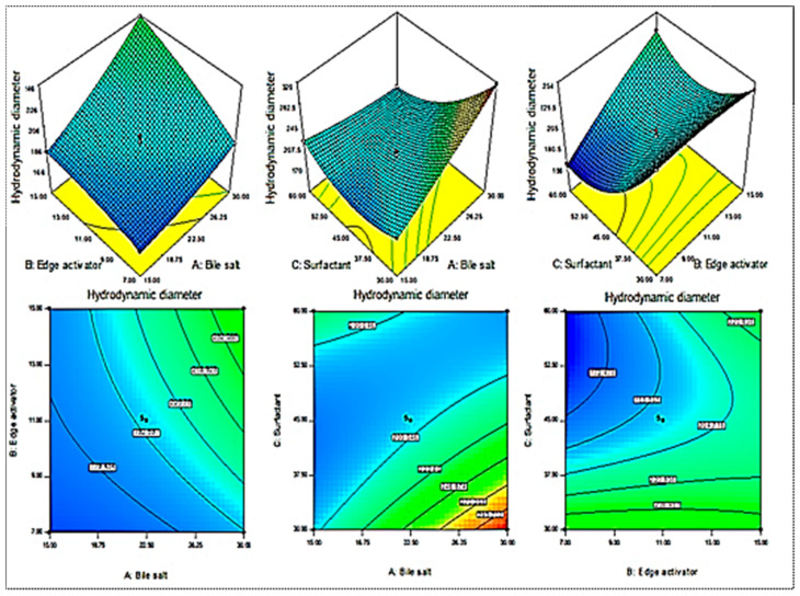
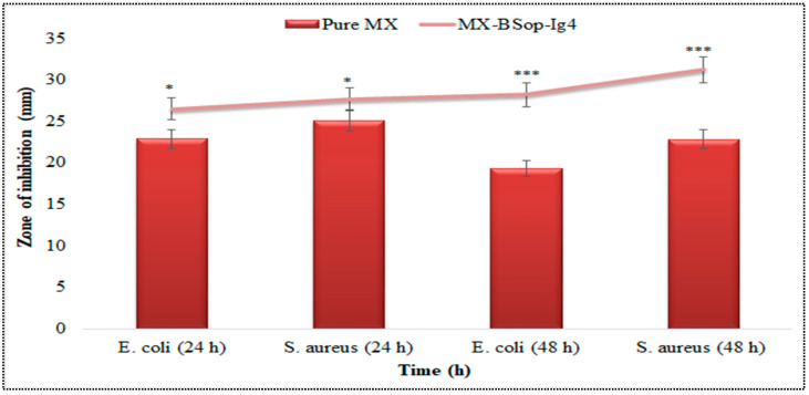
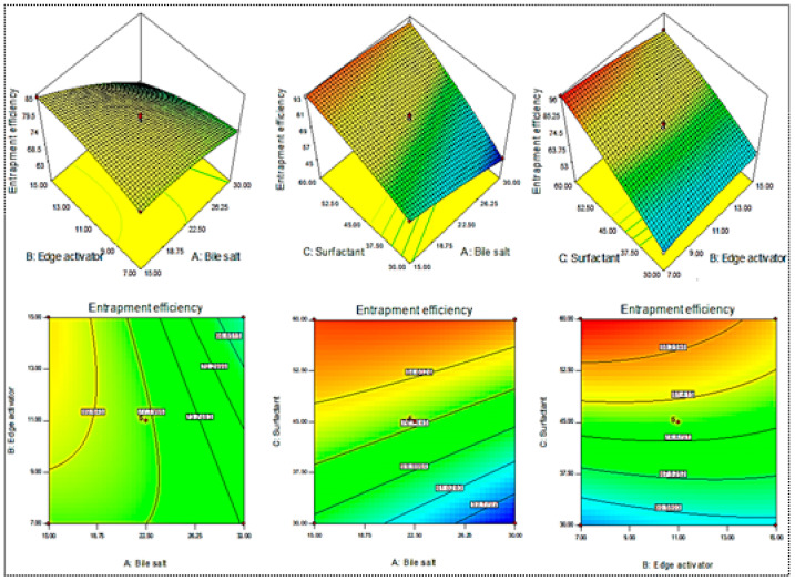
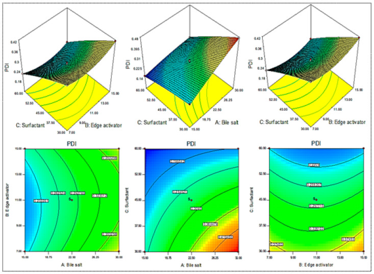
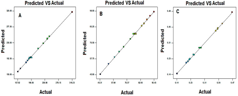
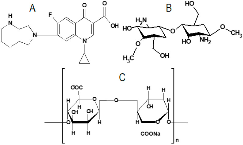
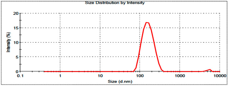
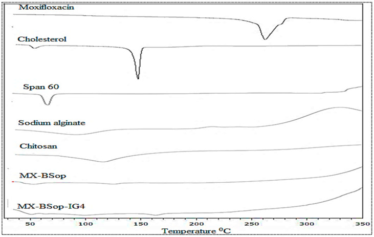
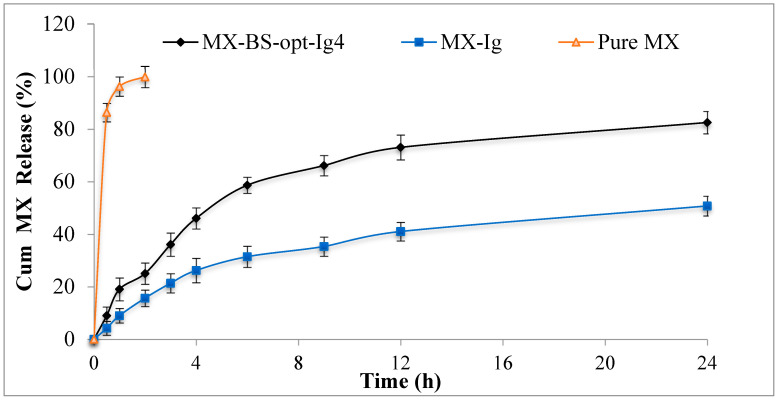
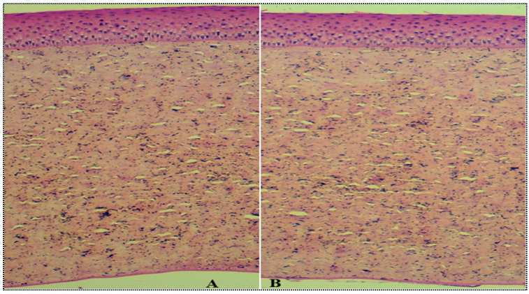
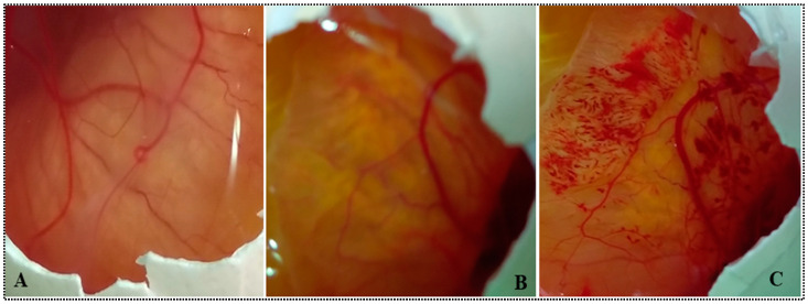
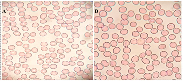
In this study, moxifloxacin (MX)-loaded bilosome (BS) in situ gel was prepared to improve ocular residence time. MX-BSs were prepared using the thin-film hydration method. They were optimized using a Box−Behnken design (BBD) with bile salt (A, sodium deoxycholate), an edge activator (B, Cremophor EL), and a surfactant (C, Span 60) as process variables. Their effects were assessed based on hydrodynamic diameter (Y1), entrapment efficacy (Y2), and polydispersity index (Y3). The optimized formulation (MX-BSop) depicted a low hydrodynamic diameter (192 ± 4 nm) and high entrapment efficiency (76 ± 1%). Further, MX-BSop was successfully transformed into an in situ gel using chitosan and sodium alginate as carriers. The optimized MX-BSop in situ gel (MX-BSop-Ig4) was further evaluated for gelling capacity, clarity, pH, viscosity, in vitro release, bio-adhesiveness, ex vivo permeation, toxicity, and antimicrobial properties. MX-BSop-Ig4 exhibited an optimum viscosity of 65.4 ± 5.3 cps in sol and 287.5 ± 10.5 cps in gel states. The sustained release profile (82 ± 4% in 24 h) was achieved with a Korsmeyer−Peppas kinetic release model (R2 = 0.9466). Significant bio-adhesion (967.9 dyne/cm2) was achieved in tear film. It also exhibited 1.2-fold and 2.8-fold higher permeation than MX-Ig and a pure MX solution, respectively. It did not show any toxicity to the tested tissue, confirmed by corneal hydration (77.3%), cornea histopathology (no internal changes), and a HET-CAM test (zero score). MX-BSop-Ig4 exhibited a significantly (p < 0.05) higher antimicrobial effect than pure MX against Staphylococcus aureus and Escherichia coli. The findings suggest that bilosome in situ gel is a good alternative to increase corneal residence time, as well as to improve therapeutic activity.
在本研究中,制备了载有莫西沙星(MX)的双分子层脂质体(BS)原位凝胶以延长眼部滞留时间。采用薄膜水化法制备MX - BS。以胆盐(A,脱氧胆酸钠)、边缘活化剂(B,聚氧乙烯蓖麻油EL)和表面活性剂(C,司盘60)作为工艺变量,使用Box - Behnken设计(BBD)对其进行优化。根据流体动力学直径(Y1)、包封率(Y2)和多分散指数(Y3)评估它们的效果。优化后的制剂(MX - BSop)呈现出较低的流体动力学直径(192±4 nm)和较高的包封效率(76±1%)。此外,以壳聚糖和海藻酸钠为载体,成功将MX - BSop转化为原位凝胶。对优化后的MX - BSop原位凝胶(MX - BSop - Ig4)的凝胶化能力、澄清度、pH值、粘度、体外释放、生物粘附性、离体渗透、毒性和抗菌性能进行了进一步评估。MX - BSop - Ig4在溶胶状态下的最佳粘度为65.4±5.3 cps,在凝胶状态下为287.5±10.5 cps。采用Korsmeyer - Peppas动力学释放模型实现了缓释曲线(24小时内释放82±4%)(R2 = 0.9466)。在泪膜中实现了显著的生物粘附(967.9达因/平方厘米)。与MX - Ig和纯MX溶液相比,其渗透率分别高出1.2倍和2.8倍。通过角膜水化(77.3%)、角膜组织病理学(无内部变化)和鸡胚绒毛尿囊膜试验(得分为零)证实,它对受试组织未显示任何毒性。与纯MX相比,MX - BSop - Ig4对金黄色葡萄球菌和大肠杆菌表现出显著更高(p < 0.05)的抗菌效果。研究结果表明,双分子层脂质体原位凝胶是延长角膜滞留时间以及提高治疗活性的良好替代品。