AO Research Institute Davos, 7270 Davos, Switzerland.
Department of Orthopedic and Trauma Surgery, University Hospital Basel, 4031 Basel, Switzerland.
Medicina (Kaunas). 2022 Jul 26;58(8):998. doi: 10.3390/medicina58080998.
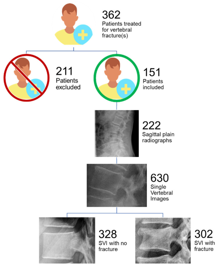
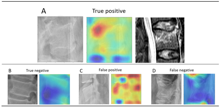
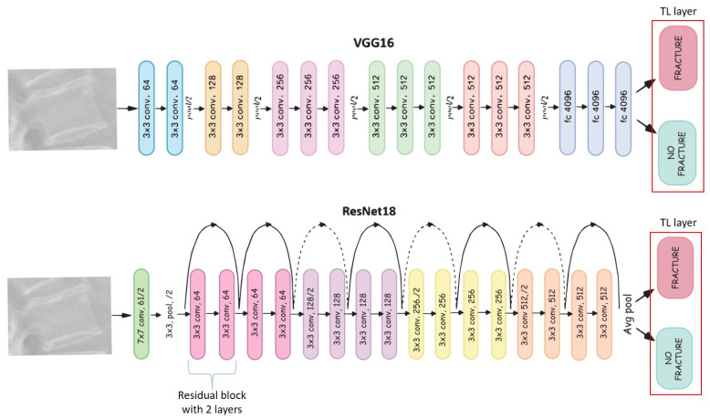
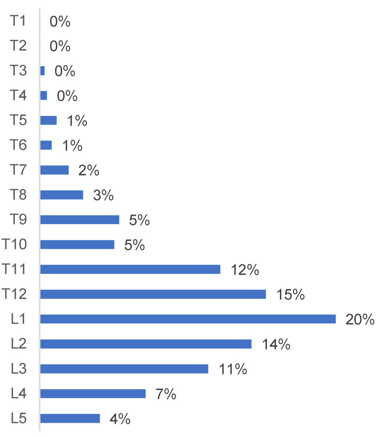
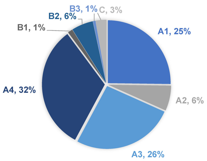
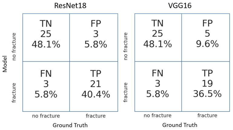
: Commonly being the first step in trauma routine imaging, up to 67% fractures are missed on plain radiographs of the thoracolumbar (TL) spine. The aim of this study was to develop a deep learning model that detects traumatic fractures on sagittal radiographs of the TL spine. Identifying vertebral fractures in simple radiographic projections would have a significant clinical and financial impact, especially for low- and middle-income countries where computed tomography (CT) and magnetic resonance imaging (MRI) are not readily available and could help select patients that need second level imaging, thus improving the cost-effectiveness. : Imaging studies (radiographs, CT, and/or MRI) of 151 patients were used. An expert group of three spinal surgeons reviewed all available images to confirm presence and type of fractures. In total, 630 single vertebra images were extracted from the sagittal radiographs of the 151 patients-302 exhibiting a vertebral body fracture, and 328 exhibiting no fracture. Following augmentation, these single vertebra images were used to train, validate, and comparatively test two deep learning convolutional neural network models, namely ResNet18 and VGG16. A heatmap analysis was then conducted to better understand the predictions of each model. : ResNet18 demonstrated a better performance, achieving higher sensitivity (91%), specificity (89%), and accuracy (88%) compared to VGG16 (90%, 83%, 86%). In 81% of the cases, the "warm zone" in the heatmaps correlated with the findings, suggestive of fracture within the vertebral body seen in the imaging studies. Vertebras T12 to L2 were the most frequently involved, accounting for 48% of the fractures. A4, A3, and A1 were the most frequent fracture types according to the AO Spine Classification. : ResNet18 could accurately identify the traumatic vertebral fractures on the TL sagittal radiographs. In most cases, the model based its prediction on the same areas that human expert classifiers used to determine the presence of a fracture.
: 通常是创伤常规成像的第一步,高达 67%的胸腰椎 (TL) 脊柱骨折在普通 X 线片上漏诊。本研究的目的是开发一种深度学习模型,用于检测 TL 脊柱矢状位 X 线片上的外伤性骨折。在简单的放射投影中识别椎体骨折将具有重要的临床和经济影响,特别是在计算机断层扫描 (CT) 和磁共振成像 (MRI) 不易获得的中低收入国家,这有助于选择需要二级成像的患者,从而提高成本效益。: 对 151 名患者的影像学研究 (X 线片、CT 和/或 MRI) 进行了回顾。一个由三名脊柱外科专家组成的专家组审查了所有可用的图像,以确认骨折的存在和类型。总共从 151 名患者的矢状位 X 线片中提取了 630 张单个椎体图像-302 张显示椎体骨折,328 张无骨折。经过扩充,这些单个椎体图像被用于训练、验证和比较测试两种深度学习卷积神经网络模型,即 ResNet18 和 VGG16。然后进行了热图分析,以更好地理解每个模型的预测。: ResNet18 表现出更好的性能,与 VGG16 (90%、83%、86%)相比,灵敏度 (91%)、特异性 (89%)和准确性 (88%)更高。在 81%的情况下,热图中的“暖区”与影像学研究中所见的椎体骨折的发现相关。T12 到 L2 的椎体最常受累,占骨折的 48%。根据 AO 脊柱分类,A4、A3 和 A1 是最常见的骨折类型。: ResNet18 可以准确识别 TL 脊柱矢状位 X 线片上的外伤性椎体骨折。在大多数情况下,该模型基于其预测与人类专家分类器用来确定骨折存在的相同区域。