• 文献检索
  • 文档翻译
  • 深度研究
  • 学术资讯
  • Suppr Zotero 插件Zotero 插件
  • 邀请有礼
  • 套餐&价格
  • 历史记录
应用&插件
Suppr Zotero 插件Zotero 插件浏览器插件Mac 客户端Windows 客户端微信小程序
定价
高级版会员购买积分包购买API积分包
服务
文献检索文档翻译深度研究API 文档MCP 服务
关于我们
关于 Suppr公司介绍联系我们用户协议隐私条款
关注我们

Suppr 超能文献

核心技术专利:CN118964589B侵权必究
粤ICP备2023148730 号-1Suppr @ 2026

文献检索

告别复杂PubMed语法,用中文像聊天一样搜索,搜遍4000万医学文献。AI智能推荐,让科研检索更轻松。

立即免费搜索

文件翻译

保留排版,准确专业,支持PDF/Word/PPT等文件格式,支持 12+语言互译。

免费翻译文档

深度研究

AI帮你快速写综述,25分钟生成高质量综述,智能提取关键信息,辅助科研写作。

立即免费体验

一种三维打印的聚己内酯-双相磷酸钙支架与在异种无血清培养基中培养的脂肪干细胞联合用于治疗骨缺损

A Three-Dimensional Printed Polycaprolactone-Biphasic-Calcium-Phosphate Scaffold Combined with Adipose-Derived Stem Cells Cultured in Xenogeneic Serum-Free Media for the Treatment of Bone Defects.

作者信息

Supphaprasitt Woraporn, Charoenmuang Lalita, Thuaksuban Nuttawut, Sangsuwan Prawichaya, Leepong Narit, Supakanjanakanti Danaiya, Vongvatcharanon Surapong, Suwanrat Trin, Srimanok Woraluk

机构信息

Department of Oral and Maxillofacial Surgery, Faculty of Dentistry, Prince of Songkla University, Hatyai 90110, Thailand.

Department of Molecular Biotechnology and Bioinformatics, Faculty of Science, Prince of Songkla University, Hatyai 90110, Thailand.

出版信息

J Funct Biomater. 2022 Jul 15;13(3):93. doi: 10.3390/jfb13030093.

DOI:10.3390/jfb13030093
PMID:35893462
原文链接:https://pmc.ncbi.nlm.nih.gov/articles/PMC9326540/
Abstract

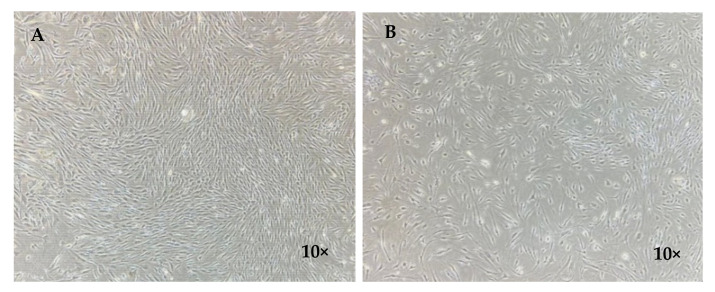
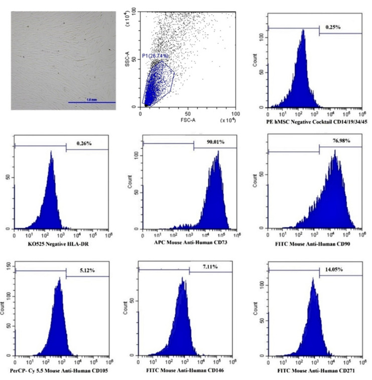
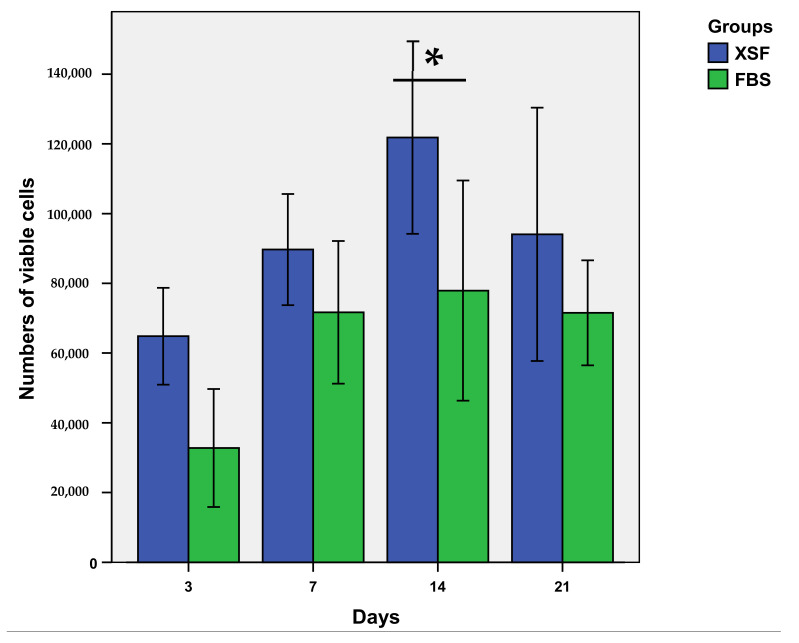
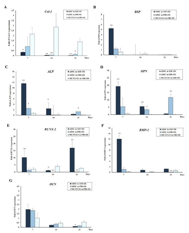
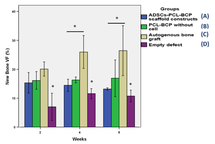
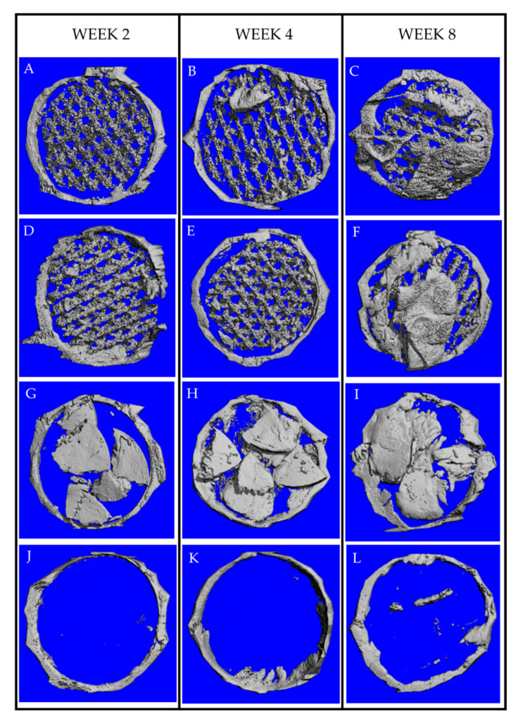
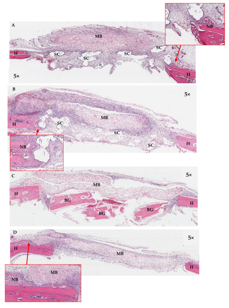
The efficacy of a three-dimensional printed polycaprolactone-biphasic-calcium-phosphate scaffold (PCL-BCP TDP scaffold) seeded with adipose-derived stem cells (ADSCs), which were cultured in xenogeneic serum-free media (XSFM) to enhance bone formation, was assessed in vitro and in animal models. The ADSCs were isolated from the buccal fat tissue of six patients using enzymatic digestion and the plastic adherence method. The proliferation and osteogenic differentiation of the cells cultured in XSFM when seeded on the scaffolds were assessed and compared with those of cells cultured in a medium containing fetal bovine serum (FBS). The cell-scaffold constructs were cultured in XSFM and were implanted into calvarial defects in thirty-six Wistar rats to assess new bone regeneration. The proliferation and osteogenic differentiation of the cells in the XSFM medium were notably better than that of the cells in the FBS medium. However, the efficacy of the constructs in enhancing new bone formation in the calvarial defects of rats was not statistically different to that achieved using the scaffolds alone. In conclusion, the PCL-BCP TDP scaffolds were biocompatible and suitable for use as an osteoconductive framework. The XSFM medium could support the proliferation and differentiation of ADSCs in vitro. However, the cell-scaffold constructs had no benefit in the enhancement of new bone formation in animal models.

摘要

评估了接种脂肪来源干细胞(ADSCs)的三维打印聚己内酯 - 双相磷酸钙支架(PCL - BCP TDP支架)的功效,这些细胞在异种无血清培养基(XSFM)中培养以促进骨形成,并在体外和动物模型中进行了评估。通过酶消化和塑料贴壁法从六名患者的颊脂肪组织中分离出ADSCs。评估了接种在支架上的细胞在XSFM中培养时的增殖和成骨分化情况,并与在含有胎牛血清(FBS)的培养基中培养的细胞进行了比较。将细胞 - 支架构建体在XSFM中培养,并植入36只Wistar大鼠的颅骨缺损处,以评估新骨再生情况。XSFM培养基中细胞的增殖和成骨分化明显优于FBS培养基中的细胞。然而,构建体在增强大鼠颅骨缺损处新骨形成方面的功效与单独使用支架所达到的功效在统计学上没有差异。总之,PCL - BCP TDP支架具有生物相容性,适合用作骨传导框架。XSFM培养基可以在体外支持ADSCs的增殖和分化。然而,细胞 - 支架构建体在动物模型中对增强新骨形成没有益处。

https://cdn.ncbi.nlm.nih.gov/pmc/blobs/130c/9326540/e8b53e1af144/jfb-13-00093-g018.jpg
https://cdn.ncbi.nlm.nih.gov/pmc/blobs/130c/9326540/78f85b3edbfd/jfb-13-00093-g001.jpg
https://cdn.ncbi.nlm.nih.gov/pmc/blobs/130c/9326540/5750a097c2d0/jfb-13-00093-g002.jpg
https://cdn.ncbi.nlm.nih.gov/pmc/blobs/130c/9326540/9515aafcd802/jfb-13-00093-g003.jpg
https://cdn.ncbi.nlm.nih.gov/pmc/blobs/130c/9326540/8342c57b1d9e/jfb-13-00093-g004.jpg
https://cdn.ncbi.nlm.nih.gov/pmc/blobs/130c/9326540/f15ec7fe1537/jfb-13-00093-g005.jpg
https://cdn.ncbi.nlm.nih.gov/pmc/blobs/130c/9326540/8e57ff355b87/jfb-13-00093-g006.jpg
https://cdn.ncbi.nlm.nih.gov/pmc/blobs/130c/9326540/f755e431d661/jfb-13-00093-g007.jpg
https://cdn.ncbi.nlm.nih.gov/pmc/blobs/130c/9326540/a6bf7eb3069e/jfb-13-00093-g008.jpg
https://cdn.ncbi.nlm.nih.gov/pmc/blobs/130c/9326540/d17601b0c702/jfb-13-00093-g009.jpg
https://cdn.ncbi.nlm.nih.gov/pmc/blobs/130c/9326540/e6e95a45bd39/jfb-13-00093-g010.jpg
https://cdn.ncbi.nlm.nih.gov/pmc/blobs/130c/9326540/5af000897e0c/jfb-13-00093-g011.jpg
https://cdn.ncbi.nlm.nih.gov/pmc/blobs/130c/9326540/0394cf36c362/jfb-13-00093-g012.jpg
https://cdn.ncbi.nlm.nih.gov/pmc/blobs/130c/9326540/a58033f0397d/jfb-13-00093-g013.jpg
https://cdn.ncbi.nlm.nih.gov/pmc/blobs/130c/9326540/d83f4741cdf0/jfb-13-00093-g014.jpg
https://cdn.ncbi.nlm.nih.gov/pmc/blobs/130c/9326540/f6fc10c2c68b/jfb-13-00093-g015.jpg
https://cdn.ncbi.nlm.nih.gov/pmc/blobs/130c/9326540/d0afac55df0d/jfb-13-00093-g016.jpg
https://cdn.ncbi.nlm.nih.gov/pmc/blobs/130c/9326540/d7b5dca5c8b4/jfb-13-00093-g017.jpg
https://cdn.ncbi.nlm.nih.gov/pmc/blobs/130c/9326540/e8b53e1af144/jfb-13-00093-g018.jpg

相似文献

1
A Three-Dimensional Printed Polycaprolactone-Biphasic-Calcium-Phosphate Scaffold Combined with Adipose-Derived Stem Cells Cultured in Xenogeneic Serum-Free Media for the Treatment of Bone Defects.一种三维打印的聚己内酯-双相磷酸钙支架与在异种无血清培养基中培养的脂肪干细胞联合用于治疗骨缺损
J Funct Biomater. 2022 Jul 15;13(3):93. doi: 10.3390/jfb13030093.
2
Effects of three-dimensionally printed polycaprolactone/β-tricalcium phosphate scaffold on osteogenic differentiation of adipose tissue- and bone marrow-derived stem cells.三维打印聚己内酯/β-磷酸三钙支架对脂肪组织和骨髓来源干细胞成骨分化的影响。
Arch Craniofac Surg. 2018 Sep;19(3):181-189. doi: 10.7181/acfs.2018.01879. Epub 2018 Sep 20.
3
Effects of polycaprolactone-biphasic calcium phosphate scaffolds on enhancing growth and differentiation of osteoblasts.聚己内酯-双相磷酸钙支架对促进成骨细胞生长和分化的影响。
Biomed Mater Eng. 2018;29(2):159-176. doi: 10.3233/BME-171720.
4
PCL/Col I-based magnetic nanocomposite scaffold provides an osteoinductive environment for ADSCs in osteogenic cues-free media conditions.基于聚己内酯/ I型胶原的磁性纳米复合支架在无成骨信号的培养基条件下为脂肪来源干细胞提供了一个骨诱导环境。
Stem Cell Res Ther. 2022 Apr 4;13(1):143. doi: 10.1186/s13287-022-02816-0.
5
Evaluation of osteoconductive effect of polycaprolactone (PCL) scaffold treated with on adipose-derived mesenchymal stem cells (ADSCs).用[具体处理方式未给出]处理的聚己内酯(PCL)支架对脂肪间充质干细胞(ADSCs)的骨传导作用评估。
Am J Stem Cells. 2023 Oct 20;12(4):83-91. eCollection 2023.
6
Evaluation of the osteogenic potential of rat adipose-derived stem cells with different polycaprolactone/alginate-based nanofibrous scaffolds: an study.不同聚己内酯/海藻酸盐基纳米纤维支架对大鼠脂肪来源干细胞成骨潜能的评估:一项研究。
Stem Cell Investig. 2020 Aug 7;7:14. doi: 10.21037/sci-2020-015. eCollection 2020.
7
[Effect of vascular endothelial growth factor 165-loaded porous poly (ε-caprolactone) scaffolds on the osteogenic differentiation of adipose-derived stem cells].[负载血管内皮生长因子165的多孔聚己内酯支架对脂肪干细胞成骨分化的影响]
Zhongguo Xiu Fu Chong Jian Wai Ke Za Zhi. 2018 Mar 15;32(3):270-275. doi: 10.7507/1002-1892.201710064.
8
Assessment of bone regeneration of a tissue-engineered bone complex using human dental pulp stem cells/poly(ε-caprolactone)-biphasic calcium phosphate scaffold constructs in rabbit calvarial defects.使用人牙髓干细胞/聚(ε-己内酯)-双相磷酸钙支架构建物评估兔颅骨缺损中组织工程骨复合物的骨再生情况。
J Mater Sci Mater Med. 2017 May;28(5):77. doi: 10.1007/s10856-017-5883-x. Epub 2017 Apr 6.
9
Stem Cell-Seeded 3D-Printed Scaffolds Combined with Self-Assembling Peptides for Bone Defect Repair.干细胞种植的 3D 打印支架与自组装肽结合用于骨缺损修复。
Tissue Eng Part A. 2022 Feb;28(3-4):111-124. doi: 10.1089/ten.TEA.2021.0055. Epub 2021 Dec 30.
10
Three-dimensional printed polycaprolactone-based scaffolds provide an advantageous environment for osteogenic differentiation of human adipose-derived stem cells.三维打印的聚己内酯基支架为人类脂肪来源干细胞的成骨分化提供了有利的环境。
J Tissue Eng Regen Med. 2018 Jan;12(1):e473-e485. doi: 10.1002/term.2310. Epub 2017 Apr 11.

引用本文的文献

1
Physical Characteristics and Biocompatibility of 3D-Printed Polylactic-Co-Glycolic Acid Membranes Used for Guided Bone Regeneration.用于引导骨再生的3D打印聚乳酸-乙醇酸共聚物膜的物理特性和生物相容性
J Funct Biomater. 2023 May 14;14(5):275. doi: 10.3390/jfb14050275.
2
IL-1b in the Secretomes of MSCs Seeded on Human Decellularized Allogeneic Bone Promotes Angiogenesis.人去细胞同种异体骨来源骨髓间充质干细胞的细胞外囊泡中的白细胞介素-1β促进血管生成。
Int J Mol Sci. 2022 Dec 4;23(23):15301. doi: 10.3390/ijms232315301.
3
Enhanced In Vitro Biocompatible Polycaprolactone/Nano-Hydroxyapatite Scaffolds with Near-Field Direct-Writing Melt Electrospinning Technology.

本文引用的文献

1
Special Features of Polyester-Based Materials for Medical Applications.用于医疗应用的聚酯基材料的特殊特性。
Polymers (Basel). 2022 Feb 27;14(5):951. doi: 10.3390/polym14050951.
2
Bioactivity of a Novel Polycaprolactone-Hydroxyapatite Scaffold Used as a Carrier of Low Dose BMP-2: An In Vitro Study.一种用作低剂量骨形态发生蛋白-2载体的新型聚己内酯-羟基磷灰石支架的生物活性:一项体外研究。
Polymers (Basel). 2021 Feb 1;13(3):466. doi: 10.3390/polym13030466.
3
Influences of Xeno-Free Media on Mesenchymal Stem Cell Expansion for Clinical Application.
采用近场直写熔体静电纺丝技术制备的具有增强体外生物相容性的聚己内酯/纳米羟基磷灰石支架
J Funct Biomater. 2022 Sep 23;13(4):161. doi: 10.3390/jfb13040161.
无血清培养基对间充质干细胞临床应用扩增的影响。
Tissue Eng Regen Med. 2021 Feb;18(1):15-23. doi: 10.1007/s13770-020-00306-z. Epub 2020 Nov 4.
4
Growth and proliferation of caprine bone marrow mesenchymal stem cells on different culture media.不同培养基上山羊骨髓间充质干细胞的生长与增殖
Tissue Cell. 2020 Dec;67:101446. doi: 10.1016/j.tice.2020.101446. Epub 2020 Oct 12.
5
Standardized xeno- and serum-free culture platform enables large-scale expansion of high-quality mesenchymal stem/stromal cells from perinatal and adult tissue sources.标准化的无动物源和无血清培养平台能够从围产期和成人组织来源大规模扩增高质量的间充质干/基质细胞。
Cytotherapy. 2021 Jan;23(1):88-99. doi: 10.1016/j.jcyt.2020.09.004. Epub 2020 Oct 21.
6
Evaluation of platelet lysate as a substitute for FBS in explant and enzymatic isolation methods of human umbilical cord MSCs.评估血小板裂解液作为人脐带间充质干细胞原代及酶消化分离法中胎牛血清的替代物。
Sci Rep. 2018 Aug 20;8(1):12439. doi: 10.1038/s41598-018-30772-4.
7
In vivo biocompatibility and degradation of novel Polycaprolactone-Biphasic Calcium phosphate scaffolds used as a bone substitute.新型聚己内酯 - 双相磷酸钙支架作为骨替代物的体内生物相容性和降解性能
Biomed Mater Eng. 2018;29(2):253-267. doi: 10.3233/BME-171727.
8
Effects of polycaprolactone-biphasic calcium phosphate scaffolds on enhancing growth and differentiation of osteoblasts.聚己内酯-双相磷酸钙支架对促进成骨细胞生长和分化的影响。
Biomed Mater Eng. 2018;29(2):159-176. doi: 10.3233/BME-171720.
9
Effect of expanded bone marrow-derived osteoprogenitor cells seeded into polycaprolactone/tricalcium phosphate scaffolds in new bone regeneration of rabbit mandibular defects.骨髓源性成骨前体细胞种植于聚己内酯/磷酸三钙支架在兔下颌骨缺损新骨再生中的作用。
J Mater Sci Mater Med. 2018 Feb 9;29(3):24. doi: 10.1007/s10856-018-6030-z.
10
Xeno-Free Strategies for Safe Human Mesenchymal Stem/Stromal Cell Expansion: Supplements and Coatings.用于安全扩增人间充质干/基质细胞的无动物源策略:补充剂和包被材料
Stem Cells Int. 2017;2017:6597815. doi: 10.1155/2017/6597815. Epub 2017 Oct 11.