• 文献检索
  • 文档翻译
  • 深度研究
  • 学术资讯
  • Suppr Zotero 插件Zotero 插件
  • 邀请有礼
  • 套餐&价格
  • 历史记录
应用&插件
Suppr Zotero 插件Zotero 插件浏览器插件Mac 客户端Windows 客户端微信小程序
定价
高级版会员购买积分包购买API积分包
服务
文献检索文档翻译深度研究API 文档MCP 服务
关于我们
关于 Suppr公司介绍联系我们用户协议隐私条款
关注我们

Suppr 超能文献

核心技术专利:CN118964589B侵权必究
粤ICP备2023148730 号-1Suppr @ 2026

文献检索

告别复杂PubMed语法,用中文像聊天一样搜索,搜遍4000万医学文献。AI智能推荐,让科研检索更轻松。

立即免费搜索

文件翻译

保留排版,准确专业,支持PDF/Word/PPT等文件格式,支持 12+语言互译。

免费翻译文档

深度研究

AI帮你快速写综述,25分钟生成高质量综述,智能提取关键信息,辅助科研写作。

立即免费体验

基于深度学习的结肠镜标本数字病理学图像的自动化组织学分类。

Automated histological classification for digital pathology images of colonoscopy specimen via deep learning.

机构信息

Department of Pathology, Hallym University Dongtan Sacred Heart Hospital, Hallym University College of Medicine, Hwaseong, Republic of Korea.

Medical Artificial Intelligence Center, Hallym University Sacred Heart Hospital, Anyang, Republic of Korea.

出版信息

Sci Rep. 2022 Jul 27;12(1):12804. doi: 10.1038/s41598-022-16885-x.

DOI:10.1038/s41598-022-16885-x
PMID:35896791
原文链接:https://pmc.ncbi.nlm.nih.gov/articles/PMC9329279/
Abstract

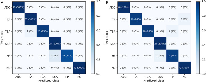
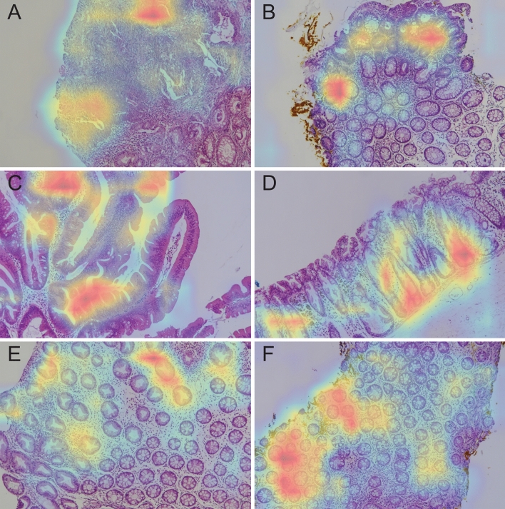
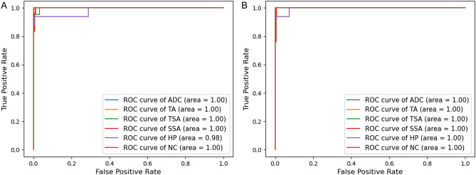
Colonoscopy is an effective tool to detect colorectal lesions and needs the support of pathological diagnosis. This study aimed to develop and validate deep learning models that automatically classify digital pathology images of colon lesions obtained from colonoscopy-related specimen. Histopathological slides of colonoscopic biopsy or resection specimens were collected and grouped into six classes by disease category: adenocarcinoma, tubular adenoma (TA), traditional serrated adenoma (TSA), sessile serrated adenoma (SSA), hyperplastic polyp (HP), and non-specific lesions. Digital photographs were taken of each pathological slide to fine-tune two pre-trained convolutional neural networks, and the model performances were evaluated. A total of 1865 images were included from 703 patients, of which 10% were used as a test dataset. For six-class classification, the mean diagnostic accuracy was 97.3% (95% confidence interval [CI], 96.0-98.6%) by DenseNet-161 and 95.9% (95% CI 94.1-97.7%) by EfficientNet-B7. The per-class area under the receiver operating characteristic curve (AUC) was highest for adenocarcinoma (1.000; 95% CI 0.999-1.000) by DenseNet-161 and TSA (1.000; 95% CI 1.000-1.000) by EfficientNet-B7. The lowest per-class AUCs were still excellent: 0.991 (95% CI 0.983-0.999) for HP by DenseNet-161 and 0.995 for SSA (95% CI 0.992-0.998) by EfficientNet-B7. Deep learning models achieved excellent performances for discriminating adenocarcinoma from non-adenocarcinoma lesions with an AUC of 0.995 or 0.998. The pathognomonic area for each class was appropriately highlighted in digital images by saliency map, particularly focusing epithelial lesions. Deep learning models might be a useful tool to help the diagnosis for pathologic slides of colonoscopy-related specimens.

摘要

结肠镜检查是一种有效的检测结直肠病变的工具,需要病理学诊断的支持。本研究旨在开发和验证深度学习模型,以便自动对结肠镜相关标本获得的结直肠病变的数字病理学图像进行分类。收集结肠镜活检或切除标本的组织病理学切片,并按疾病类别分为六类:腺癌、管状腺瘤(TA)、传统锯齿状腺瘤(TSA)、无蒂锯齿状腺瘤(SSA)、增生性息肉(HP)和非特异性病变。对每个病理切片拍摄数字照片,以微调两个预先训练的卷积神经网络,并评估模型性能。共纳入 703 名患者的 1865 张图像,其中 10%作为测试数据集。对于六类分类,DenseNet-161 的平均诊断准确率为 97.3%(95%置信区间[CI],96.0-98.6%),EfficientNet-B7 的平均诊断准确率为 95.9%(95%CI 94.1-97.7%)。DenseNet-161 对腺癌的每个类别受试者工作特征曲线(AUC)的面积最大(1.000;95%CI 0.999-1.000),EfficientNet-B7 对 TSA 的每个类别 AUC 最大(1.000;95%CI 1.000-1.000)。最低的每个类别 AUC 仍然很高:DenseNet-161 对 HP 的 AUC 为 0.991(95%CI 0.983-0.999),EfficientNet-B7 对 SSA 的 AUC 为 0.995(95%CI 0.992-0.998)。深度学习模型在区分腺癌与非腺癌病变方面表现出色,AUC 为 0.995 或 0.998。通过显著图,每个类别的特征区域在数字图像中被适当地突出显示,特别是上皮病变。深度学习模型可能是帮助诊断结肠镜相关标本的病理切片的有用工具。

https://cdn.ncbi.nlm.nih.gov/pmc/blobs/ba6a/9329279/f4d126725e69/41598_2022_16885_Fig3_HTML.jpg
https://cdn.ncbi.nlm.nih.gov/pmc/blobs/ba6a/9329279/3109bf2fbc71/41598_2022_16885_Fig1_HTML.jpg
https://cdn.ncbi.nlm.nih.gov/pmc/blobs/ba6a/9329279/40a12af2157b/41598_2022_16885_Fig2_HTML.jpg
https://cdn.ncbi.nlm.nih.gov/pmc/blobs/ba6a/9329279/f4d126725e69/41598_2022_16885_Fig3_HTML.jpg
https://cdn.ncbi.nlm.nih.gov/pmc/blobs/ba6a/9329279/3109bf2fbc71/41598_2022_16885_Fig1_HTML.jpg
https://cdn.ncbi.nlm.nih.gov/pmc/blobs/ba6a/9329279/40a12af2157b/41598_2022_16885_Fig2_HTML.jpg
https://cdn.ncbi.nlm.nih.gov/pmc/blobs/ba6a/9329279/f4d126725e69/41598_2022_16885_Fig3_HTML.jpg

相似文献

1
Automated histological classification for digital pathology images of colonoscopy specimen via deep learning.基于深度学习的结肠镜标本数字病理学图像的自动化组织学分类。
Sci Rep. 2022 Jul 27;12(1):12804. doi: 10.1038/s41598-022-16885-x.
2
Endoscopic diagnosis of sessile serrated adenoma/polyp with and without dysplasia/carcinoma.内镜诊断无蒂锯齿状腺瘤/息肉伴和不伴异型增生/癌。
World J Gastroenterol. 2018 Aug 7;24(29):3250-3259. doi: 10.3748/wjg.v24.i29.3250.
3
Impact of an Endoscopic Quality Improvement Program Focused on Adenoma Detection on Sessile Serrated Adenoma/Polyp Detection.一项聚焦腺瘤检测的内镜质量改进计划对无蒂锯齿状腺瘤/息肉检测的影响。
Dig Dis Sci. 2017 Jun;62(6):1464-1471. doi: 10.1007/s10620-017-4582-2. Epub 2017 Apr 25.
4
Automated Diagnosis of Cervical Intraepithelial Neoplasia in Histology Images via Deep Learning.通过深度学习实现组织学图像中宫颈上皮内瘤变的自动诊断
Diagnostics (Basel). 2022 Feb 21;12(2):548. doi: 10.3390/diagnostics12020548.
5
Endoscopic features of sessile serrated adenoma/polyps under narrowband imaging: A retrospective study.窄带成像内镜下无蒂锯齿状腺瘤/息肉的特征:一项回顾性研究。
J Dig Dis. 2019 Mar;20(3):135-142. doi: 10.1111/1751-2980.12706.
6
Computer-aided diagnosis of serrated colorectal lesions using non-magnified white-light endoscopic images.基于非放大白光内镜图像的结直肠锯齿状病变的计算机辅助诊断。
Int J Colorectal Dis. 2022 Aug;37(8):1875-1884. doi: 10.1007/s00384-022-04210-x. Epub 2022 Jul 21.
7
Colonoscopic features and malignant potential of sessile serrated adenomas: comparison with other serrated lesions and conventional adenomas.结肠镜下表现及无蒂锯齿状腺瘤的恶性潜能:与其他锯齿状病变和传统腺瘤的比较。
Colorectal Dis. 2016 Aug;18(8):795-802. doi: 10.1111/codi.13276.
8
Variation in the detection of serrated polyps in an average risk colorectal cancer screening cohort.在平均风险结直肠癌筛查队列中锯齿状息肉的检出存在差异。
Am J Gastroenterol. 2010 Dec;105(12):2656-64. doi: 10.1038/ajg.2010.315. Epub 2010 Aug 17.
9
Prediction of the histology of colorectal neoplasm in white light colonoscopic images using deep learning algorithms.使用深度学习算法预测白光结肠镜图像中结直肠肿瘤的组织学特征。
Sci Rep. 2021 Mar 5;11(1):5311. doi: 10.1038/s41598-021-84299-2.
10
Automated Classification of Colorectal Neoplasms in White-Light Colonoscopy Images via Deep Learning.通过深度学习实现白光结肠镜图像中大肠肿瘤的自动分类
J Clin Med. 2020 May 24;9(5):1593. doi: 10.3390/jcm9051593.

引用本文的文献

1
Deep learning model for grading carcinoma with Gini-based feature selection and linear production-inspired feature fusion.基于基尼系数的特征选择和线性生成启发式特征融合的癌症分级深度学习模型。
Sci Rep. 2025 Jul 1;15(1):21225. doi: 10.1038/s41598-025-00217-w.
2
Spatial integration of multi-omics data from serial sections using the novel Multi-Omics Imaging Integration Toolset.使用新型多组学成像整合工具集对连续切片的多组学数据进行空间整合。
Gigascience. 2025 Jan 6;14. doi: 10.1093/gigascience/giaf035.
3
Explainability of a Deep Learning-Based Classification Model for Antineutrophil Cytoplasmic Autoantibody-Associated Glomerulonephritis.

本文引用的文献

1
Artificial intelligence and computational pathology.人工智能与计算病理学。
Lab Invest. 2021 Apr;101(4):412-422. doi: 10.1038/s41374-020-00514-0. Epub 2021 Jan 16.
2
Artificial Intelligence in Health Care: Current Applications and Issues.人工智能在医疗保健中的应用及问题
J Korean Med Sci. 2020 Nov 2;35(42):e379. doi: 10.3346/jkms.2020.35.e379.
3
Whole Slide Imaging (WSI) in Pathology: Current Perspectives and Future Directions.病理全切片成像(WSI):现状与未来方向。
基于深度学习的抗中性粒细胞胞浆自身抗体相关性肾小球肾炎分类模型的可解释性
Kidney Int Rep. 2024 Nov 14;10(2):457-465. doi: 10.1016/j.ekir.2024.11.005. eCollection 2025 Feb.
4
Evaluating ChatGPT's diagnostic potential for pathology images.评估ChatGPT对病理图像的诊断潜力。
Front Med (Lausanne). 2025 Jan 23;11:1507203. doi: 10.3389/fmed.2024.1507203. eCollection 2024.
5
Deep learning model to diagnose cardiac amyloidosis from haematoxylin/eosin-stained myocardial tissue.用于从苏木精/伊红染色的心肌组织诊断心脏淀粉样变性的深度学习模型。
Eur Heart J Imaging Methods Pract. 2024 Dec 30;3(1):qyae141. doi: 10.1093/ehjimp/qyae141. eCollection 2025 Jan.
6
Improving performance in colorectal cancer histology decomposition using deep and ensemble machine learning.使用深度和集成机器学习提高结直肠癌组织学分解的性能。
Heliyon. 2024 Sep 10;10(18):e37561. doi: 10.1016/j.heliyon.2024.e37561. eCollection 2024 Sep 30.
7
Estimating infant age from skull X-ray images using deep learning.利用深度学习估算颅骨 X 光图像中的婴儿年龄。
Sci Rep. 2024 Jul 18;14(1):16600. doi: 10.1038/s41598-024-64489-4.
8
Artificial intelligence in colonoscopy: from detection to diagnosis.结肠镜检查中的人工智能:从检测到诊断。
Korean J Intern Med. 2024 Jul;39(4):555-562. doi: 10.3904/kjim.2023.332. Epub 2024 May 2.
9
Breaking Barriers: AI's Influence on Pathology and Oncology in Resource-Scarce Medical Systems.突破障碍:人工智能在资源匮乏医疗系统中对病理学和肿瘤学的影响
Cancers (Basel). 2023 Dec 2;15(23):5692. doi: 10.3390/cancers15235692.
10
Breast cancer histopathology image-based gene expression prediction using spatial transcriptomics data and deep learning.基于空间转录组学数据和深度学习的乳腺癌组织病理学图像基因表达预测。
Sci Rep. 2023 Aug 21;13(1):13604. doi: 10.1038/s41598-023-40219-0.
J Digit Imaging. 2020 Aug;33(4):1034-1040. doi: 10.1007/s10278-020-00351-z.
4
Automated Classification of Colorectal Neoplasms in White-Light Colonoscopy Images via Deep Learning.通过深度学习实现白光结肠镜图像中大肠肿瘤的自动分类
J Clin Med. 2020 May 24;9(5):1593. doi: 10.3390/jcm9051593.
5
Accuracy of artificial intelligence on histology prediction and detection of colorectal polyps: a systematic review and meta-analysis.人工智能在组织学预测和结直肠息肉检测中的准确性:系统评价和荟萃分析。
Gastrointest Endosc. 2020 Jul;92(1):11-22.e6. doi: 10.1016/j.gie.2020.02.033. Epub 2020 Feb 29.
6
Deep Learning Models for Histopathological Classification of Gastric and Colonic Epithelial Tumours.深度学习模型在胃和结肠上皮肿瘤的组织病理学分类中的应用。
Sci Rep. 2020 Jan 30;10(1):1504. doi: 10.1038/s41598-020-58467-9.
7
Single-cell transcriptome analysis reveals differential nutrient absorption functions in human intestine.单细胞转录组分析揭示了人类肠道中不同的营养吸收功能。
J Exp Med. 2020 Feb 3;217(2). doi: 10.1084/jem.20191130.
8
Genomic and transcriptomic profiling of carcinogenesis in patients with familial adenomatous polyposis.家族性腺瘤性息肉病患者癌变的基因组和转录组分析。
Gut. 2020 Jul;69(7):1283-1293. doi: 10.1136/gutjnl-2019-319438. Epub 2019 Nov 19.
9
Colonoscopy and Sigmoidoscopy Use among the Average-Risk Population for Colorectal Cancer: A Systematic Review and Trend Analysis.结肠镜检查和乙状结肠镜检查在结直肠癌平均风险人群中的应用:系统评价和趋势分析。
Cancer Prev Res (Phila). 2019 Sep;12(9):617-630. doi: 10.1158/1940-6207.CAPR-19-0202. Epub 2019 Jul 9.
10
Trends in the US and Canadian Pathologist Workforces From 2007 to 2017.2007 年至 2017 年美国和加拿大病理学家劳动力趋势。
JAMA Netw Open. 2019 May 3;2(5):e194337. doi: 10.1001/jamanetworkopen.2019.4337.