Hofmann Linda, Medyany Valentin, Ezić Jasmin, Lotfi Ramin, Niesler Beate, Röth Ralph, Engelhardt Daphne, Laban Simon, Schuler Patrick J, Hoffmann Thomas K, Brunner Cornelia, Jackson Edwin K, Theodoraki Marie-Nicole
Department of Otorhinolaryngology, Head and Neck Surgery, Ulm University Medical Center, Ulm, Germany.
Institute for Clinical Transfusion Medicine and Immunogenetics Ulm, German Red Cross Blood Services Baden-Württemberg-Hessen, Ulm, Germany.
Front Med (Lausanne). 2022 Jul 11;9:904295. doi: 10.3389/fmed.2022.904295. eCollection 2022.
Exosomes contribute to immunosuppression in head and neck squamous cell carcinoma (HNSCC), a tumor entity which lacks specific tumor biomarkers. Plasma-derived exosomes from HNSCC patients correlate with clinical parameters and have potential as liquid biopsy. Here, we investigate the cargo and functional profile of saliva-derived exosomes from HNSCC patients and their potential as non-invasive biomarkers for disease detection and immunomodulation.
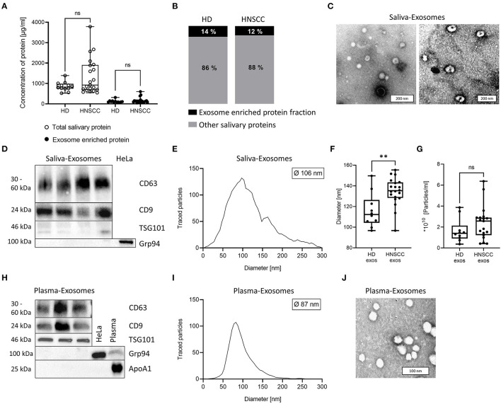
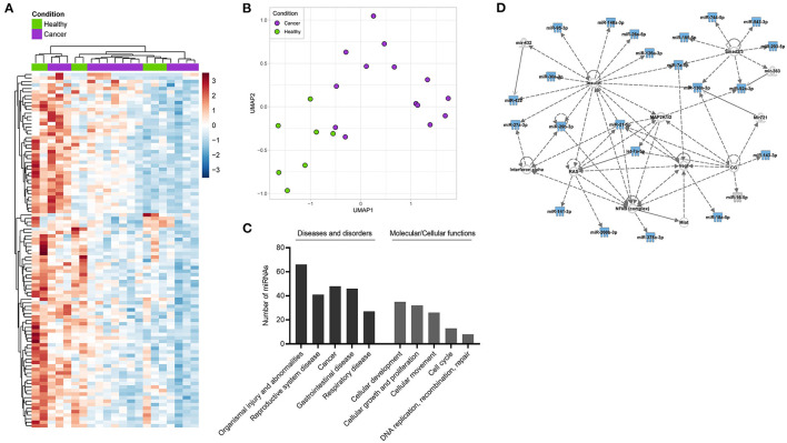
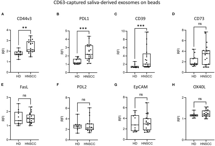
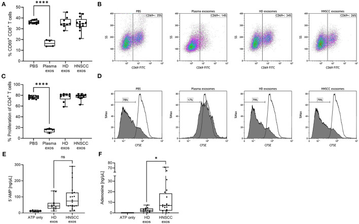
Exosomes were isolated from saliva of HNSCC patients ( = 21) and healthy donors (HD, = 12) by differential ultracentrifugation. Surface values of immune checkpoints and tumor associated antigens on saliva-derived exosomes were analyzed by bead-based flow cytometry using CD63 capture. Upon co-incubation with saliva-derived exosomes, activity and proliferation of T cells were assessed by flow cytometry (CD69 expression, CFSE assay). Adenosine levels were measured by mass spectrometry after incubation of saliva-derived exosomes with exogenous ATP. miRNA profiling of saliva-derived exosomes was performed using the nCounter SPRINT system.
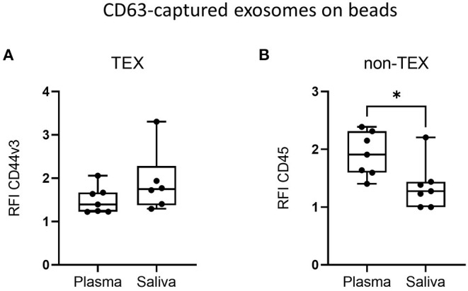
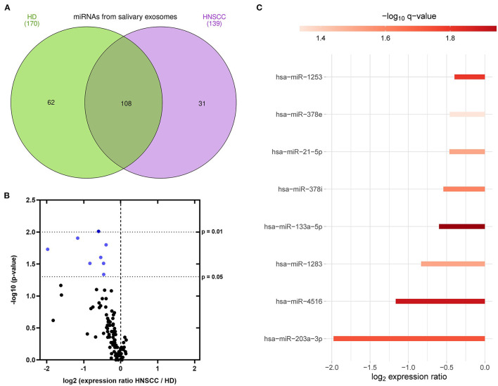
Saliva-derived, CD63-captured exosomes from HNSCC patients carried high amounts of CD44v3, PDL1 and CD39. Compared to plasma, saliva was rich in tumor-derived, CD44v3 exosomes and poor in hematopoietic cell-derived, CD45 exosomes. CD8 T cell activity was attenuated by saliva-derived exosomes from HNSCC patients, while proliferation of CD4 T cells was not affected. Further, saliva-derived exosomes produced high levels of immunosuppressive adenosine. 62 HD- and 31 HNSCC-exclusive miRNAs were identified. Samples were grouped in "Healthy" and "Cancer" based on their saliva-derived exosomal miRNA profile, which was further found to be involved in RAS/MAPK, NF-κB complex, Smad2/3, and IFN-α signaling.
Saliva-derived exosomes from HNSCC patients were enriched in tumor-derived exosomes whose cargo and functional profile reflected an immunosuppressive TME. Surface values of CD44v3, PDL1 and CD39 on CD63-captured exosomes, adenosine production and the miRNA cargo of saliva-derived exosomes emerged as discriminators of disease and emphasized their potential as liquid biomarkers specific for HNSCC.
外泌体有助于头颈部鳞状细胞癌(HNSCC)的免疫抑制,这是一种缺乏特异性肿瘤生物标志物的肿瘤实体。HNSCC患者血浆来源的外泌体与临床参数相关,具有作为液体活检的潜力。在此,我们研究HNSCC患者唾液来源外泌体的货物和功能谱及其作为疾病检测和免疫调节的非侵入性生物标志物的潜力。
通过差速超速离心从HNSCC患者(n = 21)和健康供体(HD,n = 12)的唾液中分离外泌体。使用基于磁珠的流式细胞术,通过CD63捕获分析唾液来源外泌体上免疫检查点和肿瘤相关抗原的表面值。与唾液来源的外泌体共孵育后,通过流式细胞术(CD69表达,CFSE测定)评估T细胞的活性和增殖。将唾液来源的外泌体与外源性ATP孵育后,通过质谱法测量腺苷水平。使用nCounter SPRINT系统对唾液来源的外泌体进行miRNA谱分析。
HNSCC患者唾液来源的、通过CD63捕获的外泌体携带大量CD44v3、PDL1和CD39。与血浆相比,唾液中富含肿瘤来源的CD44v3外泌体,而造血细胞来源的CD45外泌体较少。HNSCC患者唾液来源的外泌体减弱了CD8 T细胞的活性,而CD4 T细胞的增殖未受影响。此外,唾液来源的外泌体产生高水平的免疫抑制性腺苷。鉴定出62种HD特异性和31种HNSCC特异性miRNA。根据唾液来源的外泌体miRNA谱将样本分为“健康”和“癌症”两组,进一步发现其参与RAS/MAPK、NF-κB复合物、Smad2/3和IFN-α信号传导。
HNSCC患者唾液来源的外泌体富含肿瘤来源的外泌体,其货物和功能谱反映了免疫抑制性肿瘤微环境。CD63捕获的外泌体上CD44v3、PDL1和CD39的表面值、腺苷产生以及唾液来源外泌体的miRNA货物成为疾病的鉴别指标,并强调了它们作为HNSCC特异性液体生物标志物的潜力。