Department of Anesthesiology and Center for Shock, Trauma and Anesthesiology Research (STAR), University of Maryland School of Medicine, Baltimore, MD, 21201 USA.
Theranostics. 2022 Jul 11;12(12):5364-5388. doi: 10.7150/thno.72713. eCollection 2022.
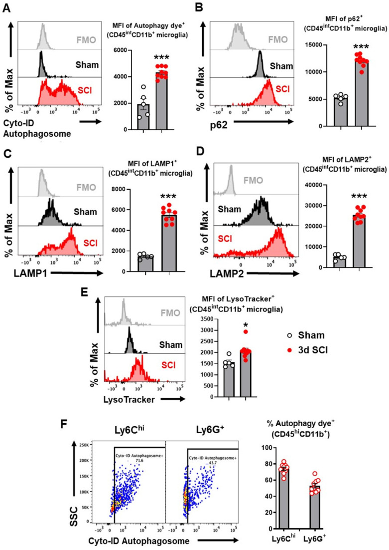
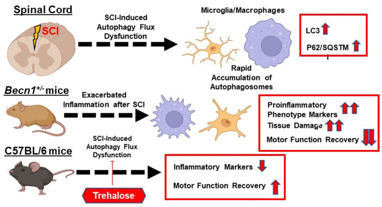
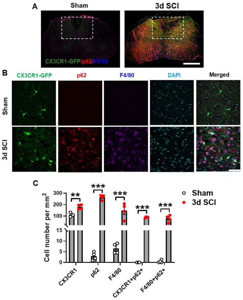
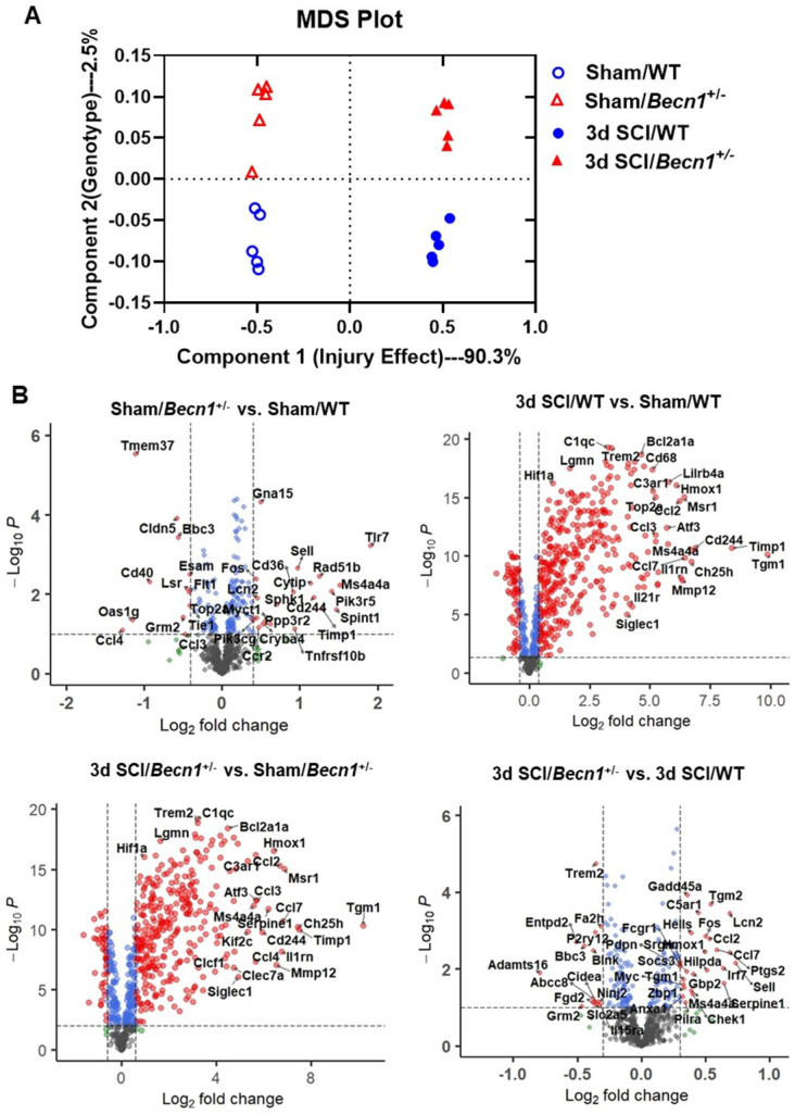
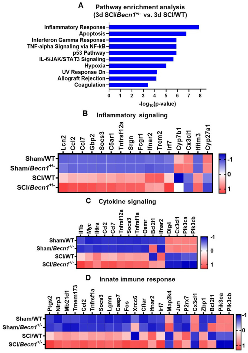
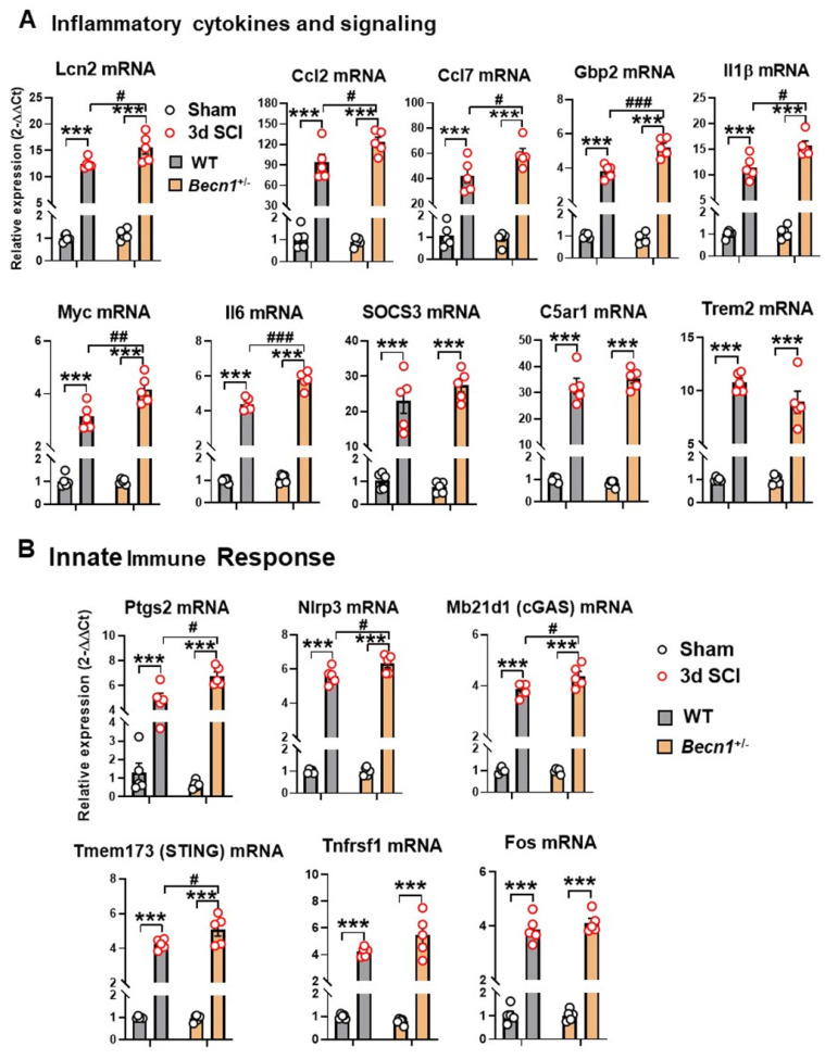
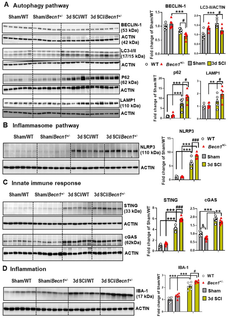
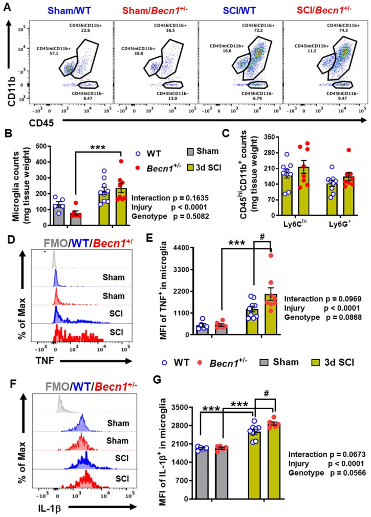
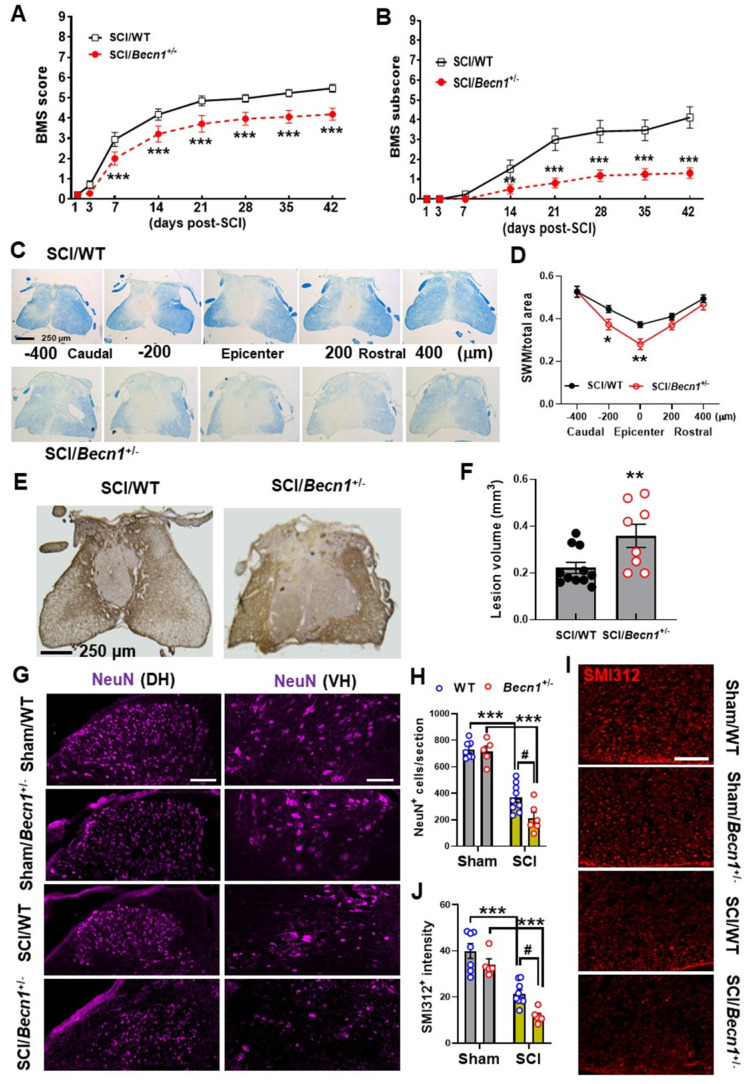
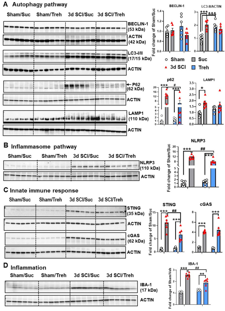
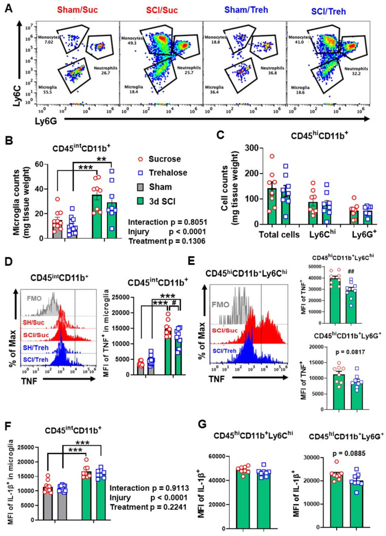
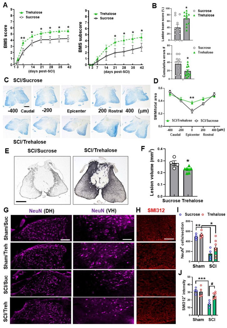
Autophagy is a catabolic process that degrades cytoplasmic constituents and organelles in the lysosome, thus serving an important role in cellular homeostasis and protection against insults. We previously reported that defects in autophagy contribute to neuronal cell damage in traumatic spinal cord injury (SCI). Recent data from other inflammatory models implicate autophagy in regulation of immune and inflammatory responses, with low levels of autophagic flux associated with pro-inflammatory phenotypes. In the present study, we examined the effects of genetically or pharmacologically manipulating autophagy on posttraumatic neuroinflammation and motor function after SCI in mice. Young adult male C57BL/6, CX3CR1-GFP, autophagy hypomorph mice, and their wildtype (WT) littermates were subjected to moderate thoracic spinal cord contusion. Neuroinflammation and autophagic flux in the injured spinal cord were assessed using flow cytometry, immunohistochemistry, and NanoString gene expression analysis. Motor function was evaluated with the Basso Mouse Scale and horizontal ladder test. Lesion volume and spared white matter were evaluated by unbiased stereology. To stimulate autophagy, disaccharide trehalose, or sucrose control, was administered in the drinking water immediately after injury and for up to 6 weeks after SCI. Flow cytometry demonstrated dysregulation of autophagic function in both microglia and infiltrating myeloid cells from the injured spinal cord at 3 days post-injury. Transgenic CX3CR1-GFP mice revealed increased autophagosome formation and inhibition of autophagic flux specifically in activated microglia/macrophages. NanoString analysis using the neuroinflammation panel demonstrated increased expression of proinflammatory genes and decreased expression of genes related to neuroprotection in mice as compared to WT controls at 3 days post-SCI. These findings were further validated by qPCR, wherein we observed significantly higher expression of proinflammatory cytokines. Western blot analysis confirmed higher protein expression of the microglia/macrophage marker IBA-1, inflammasome marker, NLRP3, and innate immune response markers cGAS and STING in mice at 3 day after SCI. Flow cytometry demonstrated that autophagy deficit did not affect either microglial or myeloid counts at 3 days post-injury, instead resulting in increased microglial production of proinflammatory cytokines. Finally, locomotor function showed significantly worse impairments in mice up to 6 weeks after SCI, which was accompanied by worsening tissue damage. Conversely, treatment with a naturally occurring autophagy inducer trehalose, reduced protein levels of p62, an adaptor protein targeting cargo to autophagosomes as well as the NLRP3, STING, and IBA-1 at 3 days post-injury. Six weeks of trehalose treatment after SCI led to improved motor function recovery as compared to control group, which was accompanied by reduced tissue damage. Our data indicate that inhibition of autophagy after SCI potentiates pro-inflammatory activation in microglia and is associated with worse functional outcomes. Conversely, increasing autophagy with trehalose, decreased inflammation and improved outcomes. These findings highlight the importance of autophagy in spinal cord microglia and its role in secondary injury after SCI.
自噬是一种溶酶体降解细胞质成分和细胞器的分解代谢过程,因此在细胞内稳态和抵抗损伤方面起着重要作用。我们之前报道过自噬缺陷导致创伤性脊髓损伤 (SCI) 中的神经元细胞损伤。最近来自其他炎症模型的数据表明自噬参与免疫和炎症反应的调节,低水平的自噬通量与促炎表型相关。在本研究中,我们研究了通过遗传或药理学方法操纵自噬对 SCI 后小鼠的创伤后神经炎症和运动功能的影响。年轻成年雄性 C57BL/6、CX3CR1-GFP、自噬缺陷型小鼠及其野生型 (WT) 同窝仔鼠接受中度胸段脊髓挫伤。通过流式细胞术、免疫组织化学和 NanoString 基因表达分析评估损伤脊髓中的神经炎症和自噬通量。使用 Basso 小鼠量表和水平梯测试评估运动功能。通过无偏立体学评估损伤体积和保留的白质。为了刺激自噬,在损伤后立即和 SCI 后 6 周内将二糖海藻糖或蔗糖对照物添加到饮用水中。流式细胞术显示,损伤后 3 天,损伤脊髓中的小胶质细胞和浸润髓样细胞的自噬功能失调。转基因 CX3CR1-GFP 小鼠显示激活的小胶质细胞/巨噬细胞中自噬体形成增加和自噬通量抑制。使用神经炎症小组的 NanoString 分析表明,与 WT 对照相比,SCI 后 3 天, 小鼠中促炎基因的表达增加,与神经保护相关的基因表达减少。qPCR 进一步验证了这一发现,其中我们观察到促炎细胞因子的表达显著增加。Western blot 分析证实,SCI 后 3 天, 小鼠中微胶质细胞/巨噬细胞标志物 IBA-1、炎症小体标志物 NLRP3 以及先天免疫反应标志物 cGAS 和 STING 的蛋白表达更高。流式细胞术表明,自噬缺陷不会影响损伤后 3 天的小胶质细胞或髓样细胞计数,反而导致小胶质细胞产生更多的促炎细胞因子。最后,运动功能显示 6 周后 SCI 的 小鼠损伤明显加重,同时组织损伤恶化。相反,用天然自噬诱导剂海藻糖治疗可降低损伤后 3 天 p62(一种将货物靶向自噬体的衔接蛋白)以及 NLRP3、STING 和 IBA-1 的蛋白水平。SCI 后 6 周的海藻糖治疗导致运动功能恢复改善,与对照组相比,组织损伤减少。我们的数据表明,SCI 后自噬的抑制增强了小胶质细胞中的促炎激活,并与更差的功能结果相关。相反,用海藻糖增加自噬会减少炎症并改善结果。这些发现强调了自噬在脊髓小胶质细胞中的重要性及其在 SCI 后的继发性损伤中的作用。