Bailo Michele, Pecco Nicolò, Callea Marcella, Scifo Paola, Gagliardi Filippo, Presotto Luca, Bettinardi Valentino, Fallanca Federico, Mapelli Paola, Gianolli Luigi, Doglioni Claudio, Anzalone Nicoletta, Picchio Maria, Mortini Pietro, Falini Andrea, Castellano Antonella
Vita-Salute San Raffaele University, Milan, Italy.
Department of Neurosurgery and Gamma Knife Radiosurgery, IRCCS Ospedale San Raffaele, Milan, Italy.
Front Neurosci. 2022 Jul 13;16:885291. doi: 10.3389/fnins.2022.885291. eCollection 2022.
Tumor heterogeneity poses major clinical challenges in high-grade gliomas (HGGs). Quantitative radiomic analysis with spatial tumor habitat clustering represents an innovative, non-invasive approach to represent and quantify tumor microenvironment heterogeneity. To date, habitat imaging has been applied mainly on conventional magnetic resonance imaging (MRI), although virtually extendible to any imaging modality, including advanced MRI techniques such as perfusion and diffusion MRI as well as positron emission tomography (PET) imaging.
This study aims to evaluate an innovative PET and MRI approach for assessing hypoxia, perfusion, and tissue diffusion in HGGs and derive a combined map for clustering of intra-tumor heterogeneity.
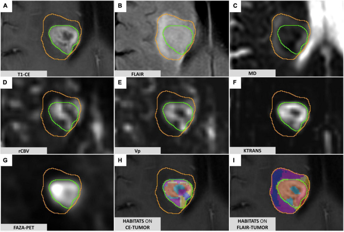
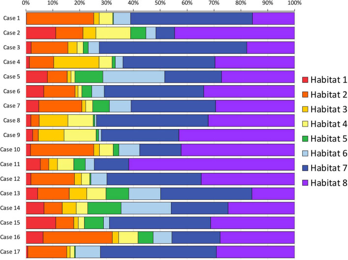
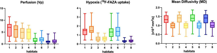
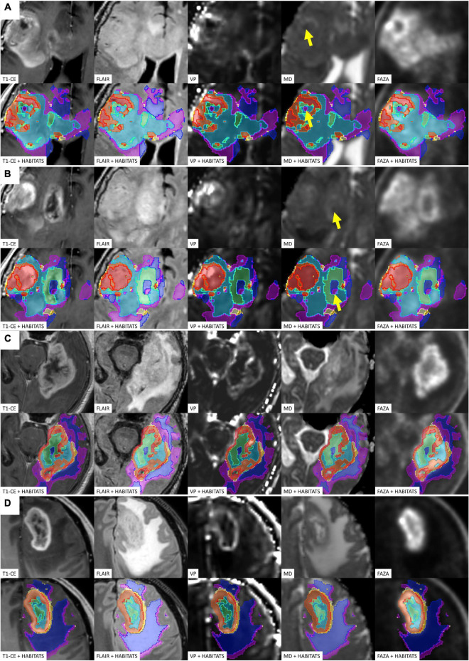
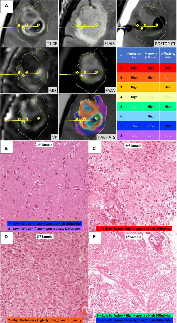
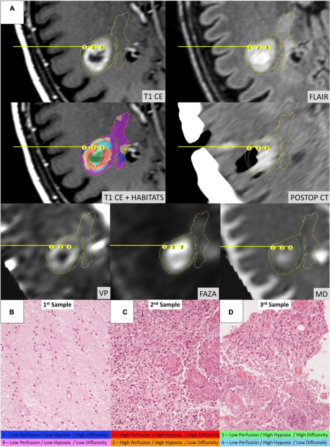
Seventeen patients harboring HGGs underwent a pre-operative acquisition of MR perfusion (PWI), Diffusion (dMRI) and F-labeled fluoroazomycinarabinoside (F-FAZA) PET imaging to evaluate tumor vascularization, cellularity, and hypoxia, respectively. Tumor volumes were segmented on fluid-attenuated inversion recovery (FLAIR) and T1 post-contrast images, and voxel-wise clustering of each quantitative imaging map identified eight combined PET and physiologic MRI habitats. Habitats' spatial distribution, quantitative features and histopathological characteristics were analyzed.
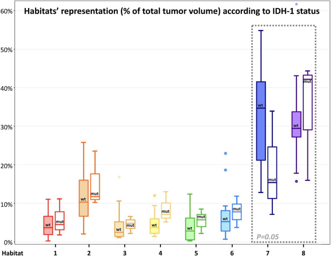
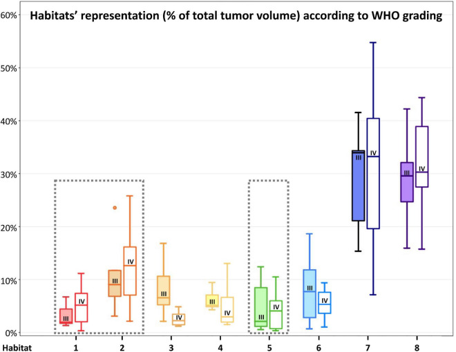
A highly reproducible distribution pattern of the clusters was observed among different cases, particularly with respect to morphological landmarks as the necrotic core, contrast-enhancing vital tumor, and peritumoral infiltration and edema, providing valuable supplementary information to conventional imaging. A preliminary analysis, performed on stereotactic bioptic samples where exact intracranial coordinates were available, identified a reliable correlation between the expected microenvironment of the different spatial habitats and the actual histopathological features. A trend toward a higher representation of the most aggressive clusters in WHO (World Health Organization) grade IV compared to WHO III was observed.
Preliminary findings demonstrated high reproducibility of the PET and MRI hypoxia, perfusion, and tissue diffusion spatial habitat maps and correlation with disease-specific histopathological features.
肿瘤异质性给高级别胶质瘤(HGGs)带来了重大临床挑战。采用空间肿瘤栖息地聚类的定量放射组学分析是一种创新的非侵入性方法,用于表征和量化肿瘤微环境异质性。迄今为止,栖息地成像主要应用于传统磁共振成像(MRI),尽管实际上可扩展到任何成像模态,包括灌注和扩散MRI等先进MRI技术以及正电子发射断层扫描(PET)成像。
本研究旨在评估一种创新的PET和MRI方法,用于评估HGGs中的缺氧、灌注和组织扩散,并得出用于肿瘤内异质性聚类的组合图谱。
17例患有HGGs的患者在术前接受了磁共振灌注(PWI)、扩散(dMRI)和F标记的氟阿糖胞苷(F-FAZA)PET成像,分别用于评估肿瘤血管生成、细胞密度和缺氧情况。在液体衰减反转恢复(FLAIR)和T1增强后图像上分割肿瘤体积,并对每个定量成像图谱进行体素级聚类,确定了八个PET与生理MRI组合栖息地。分析了栖息地的空间分布特征、定量特征和组织病理学特征。
在不同病例中观察到聚类具有高度可重复的分布模式,特别是相对于坏死核心、强化的活性肿瘤以及瘤周浸润和水肿等形态学标志而言,为传统成像提供了有价值的补充信息。对可获得精确颅内坐标的立体定向活检样本进行的初步分析,确定了不同空间栖息地的预期微环境与实际组织病理学特征之间存在可靠的相关性。观察到与世界卫生组织(WHO)III级相比,WHO IV级中最具侵袭性聚类的占比有升高趋势。
初步研究结果表明,PET和MRI缺氧、灌注及组织扩散空间栖息地图谱具有高度可重复性,且与疾病特异性组织病理学特征相关。