Department of Child and Adolescent Psychiatry/Psychology, Erasmus MC, University Medical Center Rotterdam, Rotterdam, the Netherlands.
Julius Center for Health Sciences and Primary Care, University Medical Center Utrecht, Utrecht University, Utrecht, the Netherlands.
JAMA Netw Open. 2022 Aug 1;5(8):e2224701. doi: 10.1001/jamanetworkopen.2022.24701.
Maternal tobacco use during pregnancy has been associated with various health consequences, including suboptimal neurodevelopment in offspring. However, the effect of prenatal exposure to maternal smoking on child brain development has yet to be elucidated.
To investigate the association between maternal smoking during pregnancy and offspring brain development in preadolescence as well as the mediating pathways.
DESIGN, SETTING, AND PARTICIPANTS: This prospective, population-based cohort study was embedded in the Generation R Study, Rotterdam, the Netherlands. The Generation R Study was launched in 2002, with follow-up ongoing. Child brain morphology was assessed at 9 to 11 years of age (ie, 10-12 years between exposure and outcome assessment). Data analysis was performed from March 1, 2021, to February 28, 2022, and at the time of manuscript revision. Participants included the singleton children of pregnant women residing in the study area with an expected date of delivery between April 1, 2002, and January 31, 2006; 2704 children with information on maternal smoking during pregnancy and structural neuroimaging at 9 to 11 years of age were included. A subsample of 784 children with data on DNA methylation at birth was examined in the mediation analysis.
Information on maternal smoking during pregnancy was collected via a questionnaire in each trimester. As a contrast, paternal smoking was assessed at recruitment.
Brain morphology, including brain volumes and surface-based cortical measures (thickness, surface area, and gyrification), was assessed with magnetic resonance imaging. For mediation analysis, DNA methylation at birth was quantified by a weighted methylation risk score.
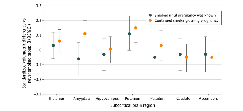
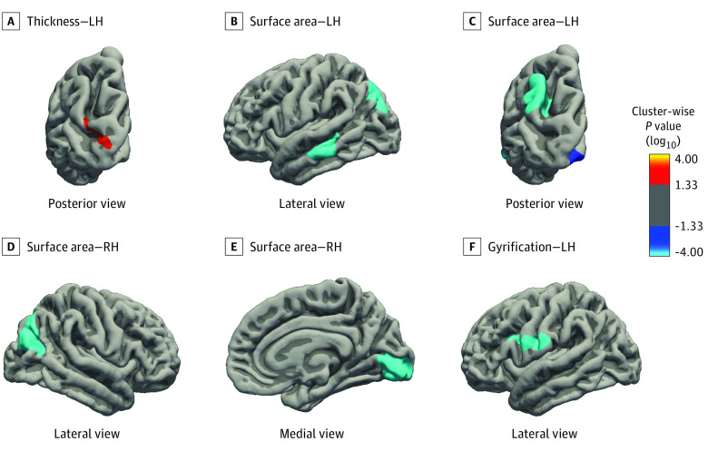
The 2704 participating children (1370 [50.7%] girls and 1334 [49.3%] boys) underwent brain imaging assessment at a mean (SD) age of 10.1 (0.6) years. Compared with nonexposed children (n = 2102), exposure to continued maternal smoking during pregnancy (n = 364) was associated with smaller total brain volume (volumetric difference [b] = -14.5 [95% CI, -25.1 to -4.0] cm3), cerebral gray matter volume (b = -7.8 [95% CI, -13.4 to -2.3] cm3), cerebral white matter volume (b = -5.9 [95% CI, -10.7 to -1.0] cm3), and surface area and less gyrification. These associations were not explained by paternal smoking nor mediated by smoking-associated DNA methylation patterns at birth. Children exposed to maternal smoking only in the first trimester (n = 238) showed no differences in brain morphology compared with nonexposed children.
The findings of this cohort study suggest that continued maternal tobacco use during pregnancy was associated with lower brain volumes and suboptimal cortical traits of offspring in preadolescence, which seemed to be independent of shared family factors. Tobacco cessation before pregnancy, or as soon as pregnancy is known, should be recommended to women for optimal brain development of their offspring.
母亲在怀孕期间吸烟与各种健康后果有关,包括后代的神经发育不良。然而,产前接触母亲吸烟对儿童大脑发育的影响尚未阐明。
研究母亲在怀孕期间吸烟与儿童在青春期前的大脑发育之间的关系以及潜在的中介途径。
设计、地点和参与者:这是一项前瞻性的、基于人群的队列研究,嵌入在荷兰鹿特丹的“生育队列研究”中。“生育队列研究”于 2002 年启动,目前仍在进行中。儿童大脑形态学在 9 至 11 岁时进行评估(即暴露和结果评估之间为 10-12 年)。数据分析于 2021 年 3 月 1 日至 2022 年 2 月 28 日进行,并在修订手稿时进行。参与者包括居住在研究区域的孕妇的单胎儿童,预计分娩日期在 2002 年 4 月 1 日至 2006 年 1 月 31 日之间;共有 2704 名儿童在 9 至 11 岁时接受了有关母亲怀孕期间吸烟和结构神经影像学的信息,包括在内。在中介分析中检查了 784 名儿童的出生时 DNA 甲基化数据。
怀孕期间母亲吸烟的信息通过每个孕期的问卷调查收集。相比之下,父亲吸烟是在招募时评估的。
脑形态学包括脑容量和基于表面的皮质测量(厚度、表面积和脑回),通过磁共振成像进行评估。对于中介分析,出生时的 DNA 甲基化通过加权甲基化风险评分进行量化。
2704 名参与儿童(1370 名女孩[50.7%]和 1334 名男孩[49.3%])在平均(标准差)年龄为 10.1(0.6)岁时接受了大脑成像评估。与未暴露儿童(n=2102)相比,怀孕期间持续接触母亲吸烟(n=364)与总脑容量较小(体积差异[b]=-14.5[95%置信区间,-25.1 至-4.0]cm3)、大脑灰质体积较小(b=-7.8[95%CI,-13.4 至-2.3]cm3)、大脑白质体积较小(b=-5.9[95%CI,-10.7 至-1.0]cm3)、表面积减少和脑回较少有关。这些关联与父亲吸烟无关,也不能由出生时与吸烟相关的 DNA 甲基化模式来解释。仅在第一孕期接触母亲吸烟的儿童(n=238)与未暴露儿童相比,大脑形态无差异。
这项队列研究的结果表明,母亲在怀孕期间持续吸烟与后代青春期前的脑容量较低和皮质特征发育不良有关,这似乎与共同的家庭因素无关。应建议女性在怀孕前或一旦知道怀孕就戒烟,以促进其子女的大脑最佳发育。