• 文献检索
  • 文档翻译
  • 深度研究
  • 学术资讯
  • Suppr Zotero 插件Zotero 插件
  • 邀请有礼
  • 套餐&价格
  • 历史记录
应用&插件
Suppr Zotero 插件Zotero 插件浏览器插件Mac 客户端Windows 客户端微信小程序
定价
高级版会员购买积分包购买API积分包
服务
文献检索文档翻译深度研究API 文档MCP 服务
关于我们
关于 Suppr公司介绍联系我们用户协议隐私条款
关注我们

Suppr 超能文献

核心技术专利:CN118964589B侵权必究
粤ICP备2023148730 号-1Suppr @ 2026

文献检索

告别复杂PubMed语法,用中文像聊天一样搜索,搜遍4000万医学文献。AI智能推荐,让科研检索更轻松。

立即免费搜索

文件翻译

保留排版,准确专业,支持PDF/Word/PPT等文件格式,支持 12+语言互译。

免费翻译文档

深度研究

AI帮你快速写综述,25分钟生成高质量综述,智能提取关键信息,辅助科研写作。

立即免费体验

卵巢囊性肿瘤的磁共振成像表现:五种在日常临床实践中常见类型的主要鉴别诊断。

Magnetic resonance imaging findings of cystic ovarian tumors: major differential diagnoses in five types frequently encountered in daily clinical practice.

机构信息

Department of Radiology, Shinshu University School of Medicine, 3-1-1 Asahi, Matsumoto, 390-8621, Japan.

出版信息

Jpn J Radiol. 2022 Dec;40(12):1213-1234. doi: 10.1007/s11604-022-01321-x. Epub 2022 Aug 2.

DOI:10.1007/s11604-022-01321-x
PMID:35916971
原文链接:https://pmc.ncbi.nlm.nih.gov/articles/PMC9719891/
Abstract

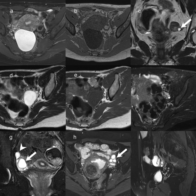
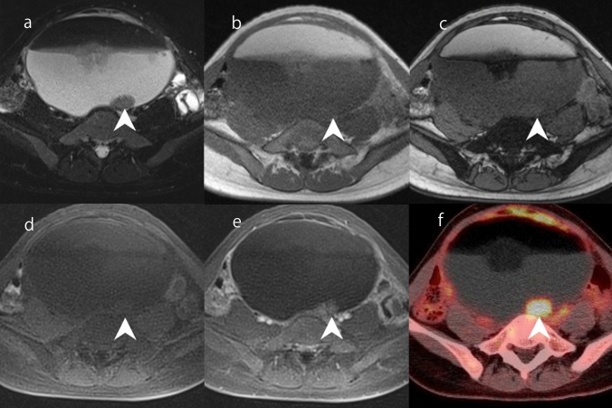
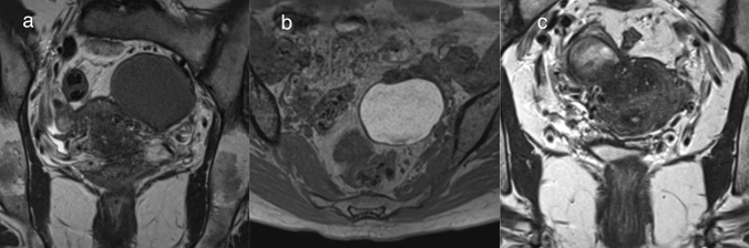
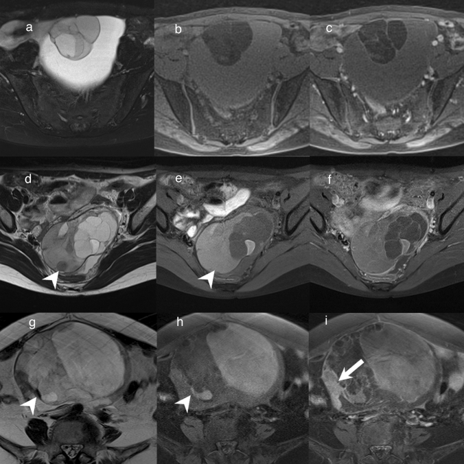
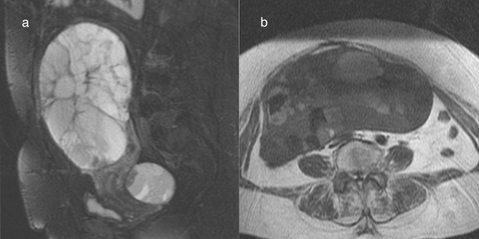
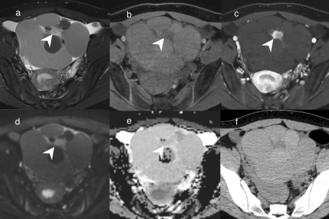
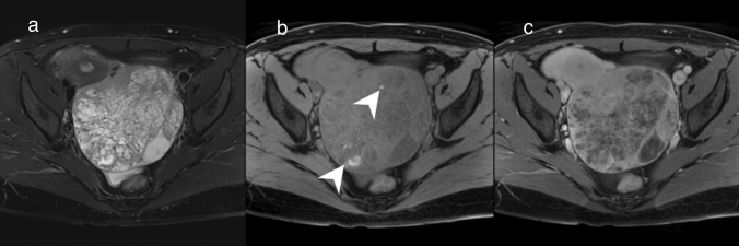
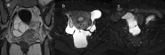
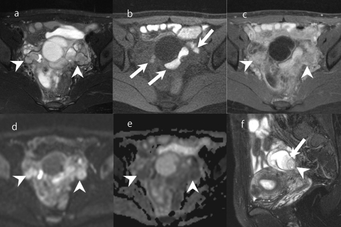
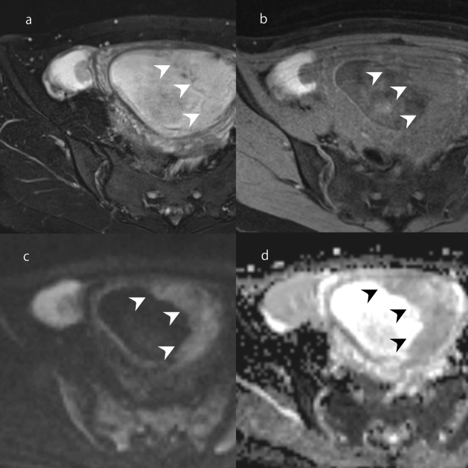
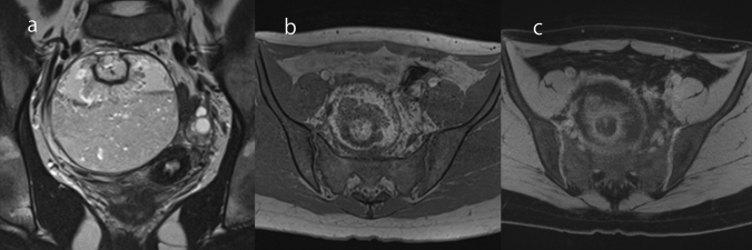
There are many types of ovarian tumors, and these different types often form cystic masses with a similar appearance, which can make their differentiation difficult. However, with the exclusion of rare ovarian tumors, the number of ovarian tumors encountered in daily practice is somewhat fixed. It goes without saying that magnetic resonance imaging (MRI) is useful for differentiating ovarian tumors. In this review, we summarize the differential diagnoses for each of the five types of MRI findings commonly encountered in daily practice. First, unilocular cystic masses without mural nodules/solid components include benign lesions such as serous cystadenoma, functional cysts, surface epithelial inclusion cysts, paratubal cysts, and endometriosis. Second, multilocular cystic ovarian lesions include mucinous tumors and ovarian metastases. It should be noted that mucinous tumors may be diagnosed as borderline or carcinoma, even if no solid component is observed. Third, cystic lesions with mural nodules that are unrelated to endometriosis include serous borderline tumor and serous carcinoma. Cystic lesions with solid components are more likely to be malignant, but some may be diagnosed as benign. Fourth, ovarian tumors deriving from endometriosis include seromucinous borderline tumors, endometrioid carcinoma, and clear cell carcinoma. These tumors sometimes need to be differentiated from serous tumors. Finally, cystic lesions with lipid contents include teratoma-related tumors. In mature cystic teratoma, mural nodules (called "Rokitansky protuberance" or "dermoid nipple") are sometimes seen, but they do not suggest malignancy. Some of these lesions can be diagnosed accurately by considering their characteristic imaging findings, their changes over time, MRI findings other than those of the primary lesion, and information from other modalities such as tumor markers. To ensure the optimal treatment for ovarian tumors, it is important to estimate the histological type as well as to diagnose whether a lesion is benign or malignant.

摘要

卵巢肿瘤有很多种类型,这些不同类型的肿瘤通常形成外观相似的囊性肿块,这使得它们的鉴别变得困难。然而,除了罕见的卵巢肿瘤外,在日常实践中遇到的卵巢肿瘤数量是相对固定的。不言而喻,磁共振成像(MRI)有助于鉴别卵巢肿瘤。在本篇综述中,我们总结了在日常实践中常见的五种 MRI 表现类型的鉴别诊断。首先,无壁结节/实性成分的单房性囊性肿块包括良性病变,如浆液性囊腺瘤、功能性囊肿、表面上皮包涵囊肿、副输卵管囊肿和子宫内膜异位症。其次,多房性囊性卵巢病变包括黏液性肿瘤和卵巢转移瘤。需要注意的是,即使没有实性成分,黏液性肿瘤也可能被诊断为交界性或癌。第三,与子宫内膜异位症无关的壁结节性囊性病变包括浆液性交界性肿瘤和浆液性癌。囊性病变伴实性成分更倾向于恶性,但有些可能被诊断为良性。第四,源于子宫内膜异位症的卵巢肿瘤包括黏液性-浆液性交界性肿瘤、子宫内膜样癌和透明细胞癌。这些肿瘤有时需要与浆液性肿瘤相鉴别。最后,含脂类物质的囊性病变包括与畸胎瘤相关的肿瘤。在成熟囊性畸胎瘤中,有时可见壁结节(称为“Rokitansky 结节”或“皮样乳头”),但这并不提示恶性。通过考虑其特征性影像学表现、随时间的变化、原发性病变以外的 MRI 表现以及肿瘤标志物等其他影像学检查的信息,可以对其中一些病变进行准确诊断。为了对卵巢肿瘤进行最佳治疗,估计组织学类型以及诊断病变是良性还是恶性是很重要的。

https://cdn.ncbi.nlm.nih.gov/pmc/blobs/d322/9719891/0d57b66a73b3/11604_2022_1321_Fig19_HTML.jpg
https://cdn.ncbi.nlm.nih.gov/pmc/blobs/d322/9719891/b0b5b6e293df/11604_2022_1321_Fig1_HTML.jpg
https://cdn.ncbi.nlm.nih.gov/pmc/blobs/d322/9719891/974ff3d2dc44/11604_2022_1321_Fig2_HTML.jpg
https://cdn.ncbi.nlm.nih.gov/pmc/blobs/d322/9719891/8df818dbd9b2/11604_2022_1321_Fig3_HTML.jpg
https://cdn.ncbi.nlm.nih.gov/pmc/blobs/d322/9719891/8f6f19b87c99/11604_2022_1321_Fig4_HTML.jpg
https://cdn.ncbi.nlm.nih.gov/pmc/blobs/d322/9719891/4b198cc96646/11604_2022_1321_Fig5_HTML.jpg
https://cdn.ncbi.nlm.nih.gov/pmc/blobs/d322/9719891/bd25313a869c/11604_2022_1321_Fig6_HTML.jpg
https://cdn.ncbi.nlm.nih.gov/pmc/blobs/d322/9719891/3202bc4199da/11604_2022_1321_Fig7_HTML.jpg
https://cdn.ncbi.nlm.nih.gov/pmc/blobs/d322/9719891/8da1a296d4f7/11604_2022_1321_Fig8_HTML.jpg
https://cdn.ncbi.nlm.nih.gov/pmc/blobs/d322/9719891/02b22208fe10/11604_2022_1321_Fig9_HTML.jpg
https://cdn.ncbi.nlm.nih.gov/pmc/blobs/d322/9719891/240e9ff8e17a/11604_2022_1321_Fig10_HTML.jpg
https://cdn.ncbi.nlm.nih.gov/pmc/blobs/d322/9719891/b654381f83f8/11604_2022_1321_Fig11_HTML.jpg
https://cdn.ncbi.nlm.nih.gov/pmc/blobs/d322/9719891/582f7036c14e/11604_2022_1321_Fig12_HTML.jpg
https://cdn.ncbi.nlm.nih.gov/pmc/blobs/d322/9719891/aad6aa44e000/11604_2022_1321_Fig13_HTML.jpg
https://cdn.ncbi.nlm.nih.gov/pmc/blobs/d322/9719891/b7a5c632844a/11604_2022_1321_Fig14_HTML.jpg
https://cdn.ncbi.nlm.nih.gov/pmc/blobs/d322/9719891/a7f651e70e98/11604_2022_1321_Fig15_HTML.jpg
https://cdn.ncbi.nlm.nih.gov/pmc/blobs/d322/9719891/10f444b1b5da/11604_2022_1321_Fig16_HTML.jpg
https://cdn.ncbi.nlm.nih.gov/pmc/blobs/d322/9719891/1e73042b93ee/11604_2022_1321_Fig17_HTML.jpg
https://cdn.ncbi.nlm.nih.gov/pmc/blobs/d322/9719891/4fc583e6ce46/11604_2022_1321_Fig18_HTML.jpg
https://cdn.ncbi.nlm.nih.gov/pmc/blobs/d322/9719891/0d57b66a73b3/11604_2022_1321_Fig19_HTML.jpg

相似文献

1
Magnetic resonance imaging findings of cystic ovarian tumors: major differential diagnoses in five types frequently encountered in daily clinical practice.卵巢囊性肿瘤的磁共振成像表现:五种在日常临床实践中常见类型的主要鉴别诊断。
Jpn J Radiol. 2022 Dec;40(12):1213-1234. doi: 10.1007/s11604-022-01321-x. Epub 2022 Aug 2.
2
Immunohistochemistry as a diagnostic aid in the evaluation of ovarian tumors.免疫组织化学作为评估卵巢肿瘤的诊断辅助手段。
Semin Diagn Pathol. 2005 Feb;22(1):3-32. doi: 10.1053/j.semdp.2005.11.002.
3
Longitudinal changes in magnetic resonance imaging of malignant and borderline tumors associated with ovarian endometriotic cyst comparing with endometriotic cysts without arising malignancy.比较与非恶性来源的卵巢子宫内膜异位囊肿相比,与恶性和交界性肿瘤相关的卵巢子宫内膜异位囊肿的磁共振成像的纵向变化。
Eur J Radiol. 2018 Aug;105:175-181. doi: 10.1016/j.ejrad.2018.06.007. Epub 2018 Jun 13.
4
Anaplastic carcinoma in ovarian seromucinous cystic tumor of borderline malignancy.交界性卵巢浆液黏液性囊腺瘤中的间变性癌。
J Ovarian Res. 2018 Sep 3;11(1):77. doi: 10.1186/s13048-018-0449-1.
5
Ultrasonic and pathological characteristics of ovarian mucinous cystic tumors with malignant mural nodules: Two cases report.伴有恶性壁结节的卵巢黏液性囊性肿瘤的超声及病理特征:两例报告
Medicine (Baltimore). 2017 Nov;96(45):e8636. doi: 10.1097/MD.0000000000008636.
6
Computed tomography of benign ovarian masses.卵巢良性肿块的计算机断层扫描
J Comput Assist Tomogr. 1985 Jul-Aug;9(4):784-9.
7
ThinPrep evaluation of fluid samples aspirated from cystic ovarian masses.对从卵巢囊性肿块中抽取的液体样本进行液基薄层制片评估。
Diagn Cytopathol. 2004 May;30(5):320-4. doi: 10.1002/dc.20046.
8
Newly Categorized Seromucinous Tumor of the Ovary: Magnetic Resonance Imaging Findings.卵巢新分类的浆液性黏液性肿瘤:磁共振成像表现
J Comput Assist Tomogr. 2019 Jan/Feb;43(1):119-127. doi: 10.1097/RCT.0000000000000776.
9
Quantitative analysis of ovarian cysts and tumors by using T2 star mapping.利用T2* 映射对卵巢囊肿和肿瘤进行定量分析。
J Obstet Gynaecol Res. 2020 Jan;46(1):140-146. doi: 10.1111/jog.14157. Epub 2019 Nov 19.
10
Germ cell tumors of the gonads: a selective review emphasizing problems in differential diagnosis, newly appreciated, and controversial issues.性腺生殖细胞肿瘤:一篇选择性综述,重点强调鉴别诊断中的问题、新认识的问题及有争议的问题。
Mod Pathol. 2005 Feb;18 Suppl 2:S61-79. doi: 10.1038/modpathol.3800310.

引用本文的文献

1
Pictorial review of bilateral adnexal lesions.双侧附件病变的影像学综述。
Abdom Radiol (NY). 2025 May 14. doi: 10.1007/s00261-025-04978-w.
2
Beautiful and regular vascularization in a borderline ovarian seromucinous tumor detected by microvascular imaging.微血管成像检测到的交界性卵巢浆液性黏液性肿瘤中美丽且规则的血管形成。
J Ovarian Res. 2025 May 2;18(1):92. doi: 10.1186/s13048-025-01675-6.
3
Three Separate Cases of Polypoid Endometriosis Coincided with Abscess, Angioleiomyoma or Stromal Sarcoma.三例息肉样子宫内膜异位症分别与脓肿、血管平滑肌瘤或间质肉瘤同时存在。

本文引用的文献

1
Ovary: MRI characterisation and O-RADS MRI.卵巢:MRI 特征及 O-RADS MRI。
Br J Radiol. 2021 Sep 1;94(1125):20210157. doi: 10.1259/bjr.20210157. Epub 2021 Apr 30.
2
Useful preoperative examination findings to classify the grade of ovarian primary mucinous tumor.有助于术前检查结果对卵巢原发性黏液性肿瘤进行分级。
Abdom Radiol (NY). 2021 Jun;46(6):2393-2402. doi: 10.1007/s00261-020-02918-4. Epub 2021 Jan 2.
3
Multimodality Imaging Approach to Ovarian Neoplasms with Pathologic Correlation.多模态影像学方法与卵巢肿瘤的病理相关性。
Int J Womens Health. 2025 Apr 13;17:1049-1054. doi: 10.2147/IJWH.S508521. eCollection 2025.
4
Two case reports of ovarian clear cell carcinoma: Atypical MRI findings with a renal corticomedullary contrast-like appearance.两例卵巢透明细胞癌病例报告:具有类似肾皮质髓质强化表现的非典型MRI表现。
Radiol Case Rep. 2025 Mar 8;20(5):2513-2520. doi: 10.1016/j.radcr.2025.01.091. eCollection 2025 May.
5
Comprehensive analysis of calcification frequency and patterns in ovarian tumours using non-contrast CT.使用非增强CT对卵巢肿瘤钙化频率和模式进行综合分析。
Jpn J Radiol. 2025 Mar 5. doi: 10.1007/s11604-025-01750-4.
6
Prediction of grading of ovarian endometrioid carcinoma using conventional MRI features.利用传统MRI特征预测卵巢子宫内膜样癌的分级
Jpn J Radiol. 2025 May;43(5):820-828. doi: 10.1007/s11604-024-01727-9. Epub 2024 Dec 28.
7
Does amide proton transfer-weighted MRI have diagnostic and differential value in ovarian cystic and predominantly cystic lesion?酰胺质子转移加权磁共振成像在卵巢囊性及以囊性为主的病变中具有诊断及鉴别价值吗?
Abdom Radiol (NY). 2024 Dec 19. doi: 10.1007/s00261-024-04768-w.
8
Teratoma combined with struma ovarii and sarcomatoid carcinoma: a case report and review of the literature.畸胎瘤合并卵巢甲状腺肿和肉瘤样癌:病例报告及文献复习。
BMC Womens Health. 2024 Sep 14;24(1):517. doi: 10.1186/s12905-024-03354-y.
9
Recent trends in AI applications for pelvic MRI: a comprehensive review.人工智能在盆腔 MRI 中的应用研究进展:综述
Radiol Med. 2024 Sep;129(9):1275-1287. doi: 10.1007/s11547-024-01861-4. Epub 2024 Aug 3.
10
Atypical Endometriosis: A Comprehensive Systematic Review of Pathological Patterns and Diagnostic Challenges.非典型子宫内膜异位症:病理模式与诊断挑战的全面系统综述
Biomedicines. 2024 May 29;12(6):1209. doi: 10.3390/biomedicines12061209.
Radiographics. 2021 Jan-Feb;41(1):289-315. doi: 10.1148/rg.2021200086. Epub 2020 Nov 13.
4
Ovarian-Adnexal Reporting Data System Magnetic Resonance Imaging (O-RADS MRI) Score for Risk Stratification of Sonographically Indeterminate Adnexal Masses.卵巢-附件报告数据系统磁共振成像(O-RADS MRI)评分用于超声不确定附件肿块的风险分层。
JAMA Netw Open. 2020 Jan 3;3(1):e1919896. doi: 10.1001/jamanetworkopen.2019.19896.
5
Apparent diffusion coefficient (ADC) values of serous, endometrioid, and clear cell carcinoma of the ovary: pathological correlation.卵巢浆液性癌、子宫内膜样癌及透明细胞癌的表观扩散系数(ADC)值:与病理的相关性
Acta Radiol. 2020 Jul;61(7):992-1000. doi: 10.1177/0284185119883392. Epub 2019 Nov 7.
6
p53, Mismatch Repair Protein, and POLE Abnormalities in Ovarian Clear Cell Carcinoma: An Outcome-based Clinicopathologic Analysis.p53、错配修复蛋白和 POLE 异常在卵巢透明细胞癌中的作用:基于结局的临床病理分析。
Am J Surg Pathol. 2019 Dec;43(12):1591-1599. doi: 10.1097/PAS.0000000000001328.
7
Trends and characteristics of epithelial ovarian cancer in Japan between 2002 and 2015: A JSGO-JSOG joint study.2002 年至 2015 年日本上皮性卵巢癌的趋势和特征:JSGO-JSOG 联合研究。
Gynecol Oncol. 2019 Jun;153(3):589-596. doi: 10.1016/j.ygyno.2019.03.243. Epub 2019 Mar 21.
8
Magnetic resonance imaging findings for discriminating clear cell carcinoma and endometrioid carcinoma of the ovary.卵巢透明细胞癌与子宫内膜样癌的磁共振成像特征鉴别。
J Ovarian Res. 2019 Feb 25;12(1):20. doi: 10.1186/s13048-019-0497-1.
9
Invasive Epithelial Ovarian Cancer Survival by Histotype and Disease Stage.不同组织学类型和疾病分期的浸润性上皮性卵巢癌生存情况。
J Natl Cancer Inst. 2019 Jan 1;111(1):60-68. doi: 10.1093/jnci/djy071.
10
Differentiation of Seromucinous Borderline Tumor from Serous Borderline Tumor on MR Imaging.磁共振成像鉴别黏液性交界性肿瘤与浆液性交界性肿瘤。
Magn Reson Med Sci. 2018 Jul 10;17(3):211-217. doi: 10.2463/mrms.mp.2017-0055. Epub 2017 Oct 6.