Kadkhodazadeh Mahdi, Fathiazar Alireza, Yadegari Zahra, Amid Reza
Dental Research Center, Research Institute of Dental Sciences, Dental School, Shahid Beheshti University of Medical Sciences, Tehran, Iran.
Department of Periodontology, Faculty of Dentistry, Ardabil University of Medical Sciences, Ardabil, Iran.
J Adv Periodontol Implant Dent. 2020 Apr 26;12(1):19-23. doi: 10.34172/japid.2020.005. eCollection 2020.
The present study aimed to evaluate the osteopromoting ability of human tooth powder and compare it to a bovine xenograft, a synthetic material, and the DFDBA allograft.
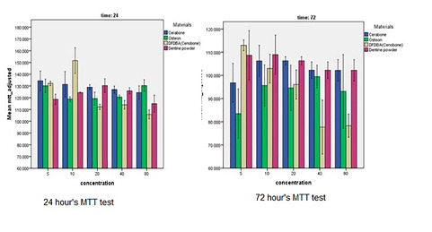
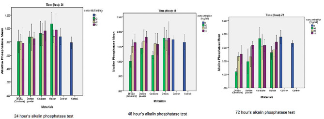
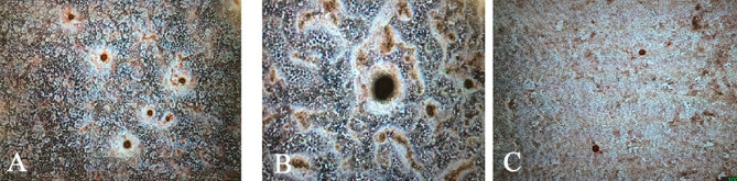
In this in vitro study, 30 teeth without caries, inflammation, and infection, which had been extracted for orthodontic reasons, were collected. The crowns were removed, pulpectomy was carried out, and the samples were ground to a powder with particles <500 µm. Osteoblast-like cells of MG-63 were cultured with the tooth powder, Cerabone, DFDBA, and Osteon II. Cell proliferation was assessed by the MTT assay at 24- and 72-hour intervals. The alizarin red test was carried out after three and five days. The alkaline phosphatase level was measured after 24, 48, and 72 hours to assess the osteoblastic activity. The results were analyzed with one-way ANOVA.
According to the MTT assay, all the materials exhibited a higher proliferation rate than the control group in 24 hours. In 72 hours, DFDBA had the lowest cell proliferation rate at concentrations of 40 and 80 mg/mL. DFDBA and the positive control group were able to create calcified nodules by the alizarin red test. At the 48- and 72-hour intervals, DFDBA had the lowest alkaline phosphatase activity at a concentration of 40 mg/mL. At the 72-hour interval, bovine xenograft had the highest alkaline phosphatase level, followed by the synthetic material and tooth powder.
The tooth powder was able to increase cell proliferation in comparison with the bovine xenograft, the synthetic graft, and the DFDBA. However, its osteopromoting ability was less than that of the osteogenic materials.
本研究旨在评估人牙粉的骨促进能力,并将其与牛异种移植物、合成材料和脱矿冻干骨同种异体移植物进行比较。
在这项体外研究中,收集了30颗因正畸原因拔除的无龋、无炎症和无感染的牙齿。去除牙冠,进行牙髓摘除术,将样本研磨成颗粒<500 µm的粉末。将MG-63成骨样细胞与牙粉、Cerabone、脱矿冻干骨和Osteon II一起培养。每隔24小时和72小时通过MTT法评估细胞增殖。在三天和五天后进行茜素红试验。在24、48和72小时后测量碱性磷酸酶水平,以评估成骨细胞活性。结果采用单因素方差分析进行分析。
根据MTT法,所有材料在24小时时的增殖率均高于对照组。在72小时时,脱矿冻干骨在浓度为40和80 mg/mL时细胞增殖率最低。通过茜素红试验,脱矿冻干骨和阳性对照组能够形成钙化结节。在48小时和72小时时,脱矿冻干骨在浓度为40 mg/mL时碱性磷酸酶活性最低。在72小时时,牛异种移植物的碱性磷酸酶水平最高,其次是合成材料和牙粉。
与人牙粉相比,牛异种移植物、合成移植物和脱矿冻干骨均能促进细胞增殖。然而,其骨促进能力低于成骨材料。