• 文献检索
  • 文档翻译
  • 深度研究
  • 学术资讯
  • Suppr Zotero 插件Zotero 插件
  • 邀请有礼
  • 套餐&价格
  • 历史记录
应用&插件
Suppr Zotero 插件Zotero 插件浏览器插件Mac 客户端Windows 客户端微信小程序
定价
高级版会员购买积分包购买API积分包
服务
文献检索文档翻译深度研究API 文档MCP 服务
关于我们
关于 Suppr公司介绍联系我们用户协议隐私条款
关注我们

Suppr 超能文献

核心技术专利:CN118964589B侵权必究
粤ICP备2023148730 号-1Suppr @ 2026

文献检索

告别复杂PubMed语法,用中文像聊天一样搜索,搜遍4000万医学文献。AI智能推荐,让科研检索更轻松。

立即免费搜索

文件翻译

保留排版,准确专业,支持PDF/Word/PPT等文件格式,支持 12+语言互译。

免费翻译文档

深度研究

AI帮你快速写综述,25分钟生成高质量综述,智能提取关键信息,辅助科研写作。

立即免费体验

基于微泡分离的短采集时间超高分辨率超声微血管成像。

Short Acquisition Time Super-Resolution Ultrasound Microvessel Imaging via Microbubble Separation.

机构信息

Department of Radiology, Mayo Clinic College of Medicine and Science, Mayo Clinic, Rochester, MN, USA.

Beckman Institute, University of Illinois at Urbana-Champaign, Urbana, IL, USA.

出版信息

Sci Rep. 2020 Apr 7;10(1):6007. doi: 10.1038/s41598-020-62898-9.

DOI:10.1038/s41598-020-62898-9
PMID:32265457
原文链接:https://pmc.ncbi.nlm.nih.gov/articles/PMC7138805/
Abstract

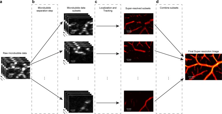
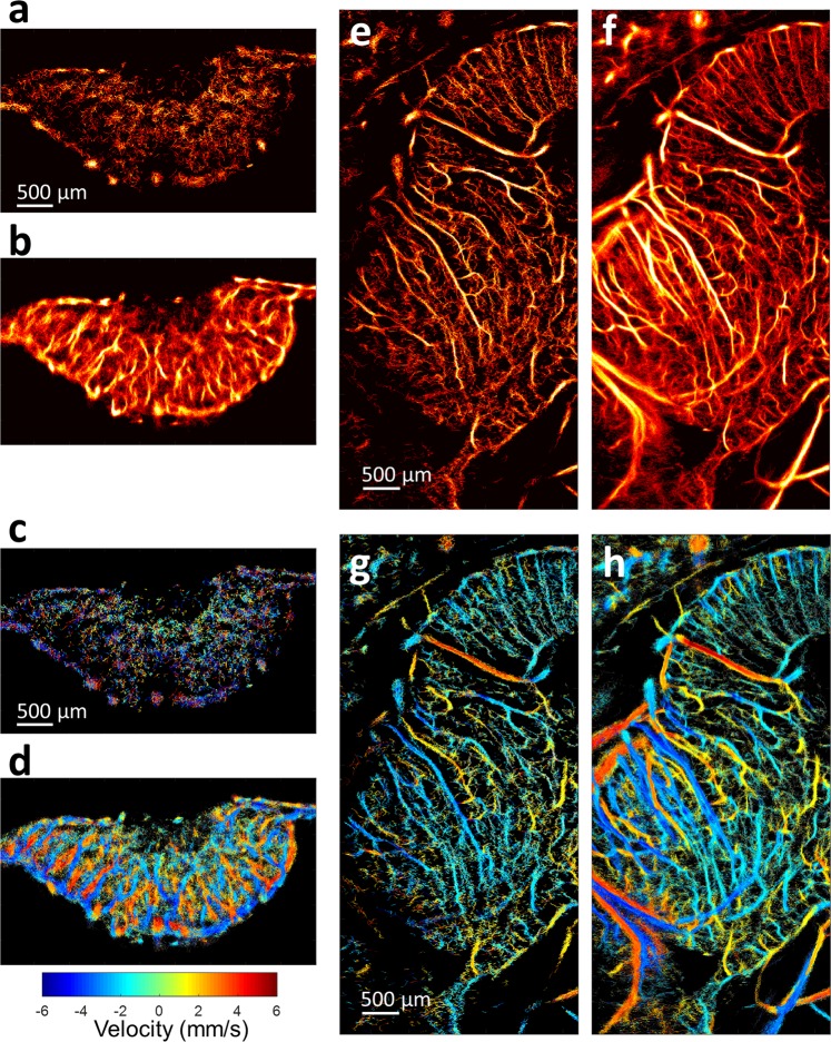
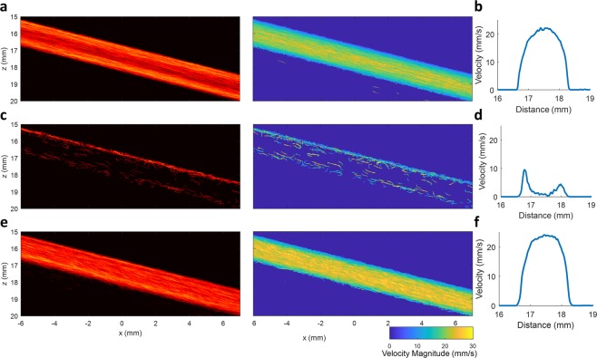
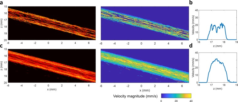
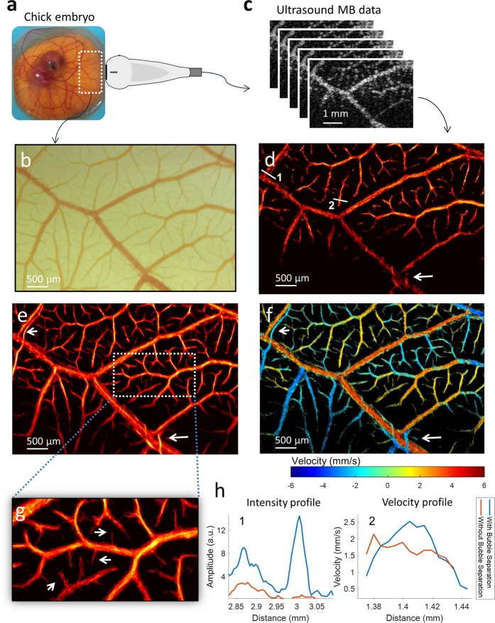
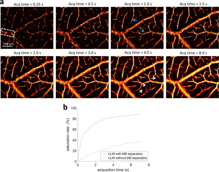
Super-resolution ultrasound localization microscopy (ULM), based on localization and tracking of individual microbubbles (MBs), offers unprecedented microvascular imaging resolution at clinically relevant penetration depths. However, ULM is currently limited by the requirement of dilute MB concentrations to ensure spatially sparse MB events for accurate localization and tracking. The corresponding long imaging acquisition times (tens of seconds or several minutes) to accumulate sufficient isolated MB events for full reconstruction of microvasculature preclude the clinical translation of the technique. To break this fundamental tradeoff between acquisition time and MB concentration, in this paper we propose to separate spatially overlapping MB events into sub-populations, each with sparser MB concentration, based on spatiotemporal differences in the flow dynamics (flow speeds and directions). MB localization and tracking are performed for each sub-population separately, permitting more robust ULM imaging of high-concentration MB injections. The superiority of the proposed MB separation technique over conventional ULM processing is demonstrated in flow channel phantom data, and in the chorioallantoic membrane of chicken embryos with optical imaging as an in vivo reference standard. Substantial improvement of ULM is further demonstrated on a chicken embryo tumor xenograft model and a chicken brain, showing both morphological and functional microvasculature details at super-resolution within a short acquisition time (several seconds). The proposed technique allows more robust MB localization and tracking at relatively high MB concentrations, alleviating the need for dilute MB injections, and thereby shortening the acquisition time of ULM imaging and showing great potential for clinical translation.

摘要

基于单个微泡(MB)定位和跟踪的超分辨率超声定位显微镜(ULM),在临床相关穿透深度下提供了前所未有的微血管成像分辨率。然而,ULM 目前受到需要稀释 MB 浓度的限制,以确保空间稀疏的 MB 事件用于准确的定位和跟踪。相应的长成像采集时间(数十秒或数分钟)来积累足够的孤立 MB 事件以完全重建微血管,排除了该技术的临床转化。为了打破采集时间和 MB 浓度之间的这种基本权衡,在本文中,我们提出了一种基于流动力学(流速和方向)的时空差异,将空间重叠的 MB 事件分离成具有更稀疏 MB 浓度的亚群。对每个亚群分别进行 MB 定位和跟踪,允许对高浓度 MB 注射进行更稳健的 ULM 成像。所提出的 MB 分离技术在流动通道幻影数据和鸡胚的脉络丛膜中表现出优于传统 ULM 处理的优越性,作为体内参考标准的光学成像。在鸡胚肿瘤异种移植模型和鸡脑中进一步证明了 ULM 的显著改进,在短采集时间(几秒钟)内以超分辨率显示了形态和功能微血管细节。该技术允许在相对较高的 MB 浓度下进行更稳健的 MB 定位和跟踪,减轻了对稀释 MB 注射的需求,从而缩短了 ULM 成像的采集时间,并显示出临床转化的巨大潜力。

https://cdn.ncbi.nlm.nih.gov/pmc/blobs/e922/7138805/22225bab8725/41598_2020_62898_Fig6_HTML.jpg
https://cdn.ncbi.nlm.nih.gov/pmc/blobs/e922/7138805/47a9de5df9ff/41598_2020_62898_Fig1_HTML.jpg
https://cdn.ncbi.nlm.nih.gov/pmc/blobs/e922/7138805/53c117338039/41598_2020_62898_Fig2_HTML.jpg
https://cdn.ncbi.nlm.nih.gov/pmc/blobs/e922/7138805/1c5e8f04f6fe/41598_2020_62898_Fig3_HTML.jpg
https://cdn.ncbi.nlm.nih.gov/pmc/blobs/e922/7138805/bf969ee24aa7/41598_2020_62898_Fig4_HTML.jpg
https://cdn.ncbi.nlm.nih.gov/pmc/blobs/e922/7138805/cefc89816825/41598_2020_62898_Fig5_HTML.jpg
https://cdn.ncbi.nlm.nih.gov/pmc/blobs/e922/7138805/22225bab8725/41598_2020_62898_Fig6_HTML.jpg

相似文献

1
Short Acquisition Time Super-Resolution Ultrasound Microvessel Imaging via Microbubble Separation.基于微泡分离的短采集时间超高分辨率超声微血管成像。
Sci Rep. 2020 Apr 7;10(1):6007. doi: 10.1038/s41598-020-62898-9.
2
Improved Ultrasound Localization Microscopy Based on Microbubble Uncoupling via Transmit Excitation.基于微泡解耦的超声定位显微镜的改进:通过发射激励。
IEEE Trans Ultrason Ferroelectr Freq Control. 2022 Mar;69(3):1041-1052. doi: 10.1109/TUFFC.2022.3143864. Epub 2022 Mar 2.
3
Fast super-resolution ultrasound microvessel imaging using spatiotemporal data with deep fully convolutional neural network.基于深度全卷积神经网络的时空域数据快速超高分辨率超声微血管成像。
Phys Med Biol. 2021 Mar 23;66(7). doi: 10.1088/1361-6560/abeb31.
4
Super-resolution ultrasound localization microscopy based on a high frame-rate clinical ultrasound scanner: an in-human feasibility study.基于高帧率临床超声扫描仪的超分辨率超声定位显微镜:在人体中的可行性研究。
Phys Med Biol. 2021 Apr 8;66(8). doi: 10.1088/1361-6560/abef45.
5
Curvelet Transform-Based Sparsity Promoting Algorithm for Fast Ultrasound Localization Microscopy.基于 Curvelet 变换的稀疏促进算法在快速超声定位显微镜中的应用。
IEEE Trans Med Imaging. 2022 Sep;41(9):2385-2398. doi: 10.1109/TMI.2022.3162839. Epub 2022 Aug 31.
6
Deep Learning-Based Microbubble Localization for Ultrasound Localization Microscopy.基于深度学习的超声定位显微镜微泡定位。
IEEE Trans Ultrason Ferroelectr Freq Control. 2022 Apr;69(4):1312-1325. doi: 10.1109/TUFFC.2022.3152225. Epub 2022 Mar 30.
7
Frame rate effects and their compensation on super-resolution microvessel imaging using ultrasound localization microscopy.帧频效应及其在超声定位显微镜超分辨微血管成像中的补偿。
Ultrasonics. 2023 Jul;132:107009. doi: 10.1016/j.ultras.2023.107009. Epub 2023 Apr 10.
8
Ultrasound localization microscopy.超声定位显微镜。
Z Med Phys. 2023 Aug;33(3):292-308. doi: 10.1016/j.zemedi.2023.02.004. Epub 2023 Jun 15.
9
Kalman Filter-Based Microbubble Tracking for Robust Super-Resolution Ultrasound Microvessel Imaging.基于卡尔曼滤波的微泡跟踪用于稳健的超声微血管超分辨率成像。
IEEE Trans Ultrason Ferroelectr Freq Control. 2020 Sep;67(9):1738-1751. doi: 10.1109/TUFFC.2020.2984384. Epub 2020 Mar 31.
10
A Forked Microvascular Phantom for Ultrasound Localization Microscopy Investigations.用于超声定位显微镜研究的分叉微血管模型。
IEEE Trans Ultrason Ferroelectr Freq Control. 2024 Jul;71(7):887-896. doi: 10.1109/TUFFC.2024.3409518. Epub 2024 Jul 9.

引用本文的文献

1
Deformable detection transformers for domain adaptable ultrasound localization microscopy with robustness to point spread function variations.用于具有对点扩散函数变化鲁棒性的域自适应超声定位显微镜的可变形检测变压器。
Sci Rep. 2025 Jul 10;15(1):24840. doi: 10.1038/s41598-025-09120-w.
2
Differentiation of benign and malignant breast lesions by ultrasound localization microscopy.超声定位显微镜对乳腺良恶性病变的鉴别诊断
Insights Imaging. 2025 Jun 18;16(1):128. doi: 10.1186/s13244-025-02013-6.
3
Progresses and clinical application of super-resolution ultrasound imaging: a narrative review.

本文引用的文献

1
Super-Resolution Ultrasound Localization Microscopy Through Deep Learning.基于深度学习的超高分辨率超声定位显微镜
IEEE Trans Med Imaging. 2021 Mar;40(3):829-839. doi: 10.1109/TMI.2020.3037790. Epub 2021 Mar 2.
2
Kalman Filter-Based Microbubble Tracking for Robust Super-Resolution Ultrasound Microvessel Imaging.基于卡尔曼滤波的微泡跟踪用于稳健的超声微血管超分辨率成像。
IEEE Trans Ultrason Ferroelectr Freq Control. 2020 Sep;67(9):1738-1751. doi: 10.1109/TUFFC.2020.2984384. Epub 2020 Mar 31.
3
Ultrasound localization microscopy of renal tumor xenografts in chicken embryo is correlated to hypoxia.
超分辨率超声成像的进展与临床应用:一篇叙述性综述
Ultrasound J. 2025 Jun 16;17(1):29. doi: 10.1186/s13089-025-00432-6.
4
Enhanced ultrasound particle image velocimetry (E-uPIV) enables fast flow mapping of microvasculature.增强型超声粒子图像测速技术(E-uPIV)能够对微血管进行快速血流成像。
Commun Eng. 2025 May 14;4(1):88. doi: 10.1038/s44172-025-00423-4.
5
Transcranial ultrasound localization microscopy in moyamoya patients using a clinical ultrasound system.使用临床超声系统对烟雾病患者进行经颅超声定位显微镜检查。
Theranostics. 2025 Mar 10;15(9):4074-4083. doi: 10.7150/thno.105427. eCollection 2025.
6
Optimizingdata acquisition for robust clinical microvascular imaging using ultrasound localization microscopy.利用超声定位显微镜优化数据采集以实现稳健的临床微血管成像。
Phys Med Biol. 2025 Mar 27;70(7). doi: 10.1088/1361-6560/adc0de.
7
Mapping of prostate cancer microvascular patterns using super-resolution ultrasound imaging.使用超分辨率超声成像绘制前列腺癌微血管模式
Eur Radiol Exp. 2025 Feb 20;9(1):25. doi: 10.1186/s41747-025-00561-6.
8
Dual-modal super-resolution ultrasound and NIR-II fluorescence imaging of ischemic stroke with ICG-doped porous PLGA microspheres.使用吲哚菁绿掺杂的多孔聚乳酸-羟基乙酸共聚物微球对缺血性中风进行双模态超分辨率超声和近红外二区荧光成像
Mater Today Bio. 2025 Jan 22;31:101513. doi: 10.1016/j.mtbio.2025.101513. eCollection 2025 Apr.
9
Ultrasound localization microscopy in the diagnosis of breast tumors and prediction of relevant histologic biomarkers associated with prognosis in humans: the protocol for a prospective, multicenter study.超声定位显微镜在人类乳腺肿瘤诊断及与预后相关的组织学生物标志物预测中的应用:一项前瞻性多中心研究方案
BMC Med Imaging. 2025 Jan 8;25(1):13. doi: 10.1186/s12880-024-01535-7.
10
Ultrasound super-resolution imaging for non-invasive assessment of microvessel in prostate lesion.超声超分辨率成像用于前列腺病变微血管的无创评估。
Cancer Imaging. 2025 Jan 7;25(1):1. doi: 10.1186/s40644-024-00819-z.
鸡胚肾肿瘤异种移植的超声定位显微镜检查与缺氧相关。
Sci Rep. 2020 Feb 12;10(1):2478. doi: 10.1038/s41598-020-59338-z.
4
3D Super-Resolution US Imaging of Rabbit Lymph Node Vasculature in Vivo by Using Microbubbles.利用微泡进行体内兔淋巴结脉管系统的 3D 超分辨率超声成像
Radiology. 2019 Jun;291(3):642-650. doi: 10.1148/radiol.2019182593. Epub 2019 Apr 16.
5
Fast Acoustic Wave Sparsely Activated Localization Microscopy (fast-AWSALM): Ultrasound Super-Resolution using Plane-Wave Activation of Nanodroplets.快速声波稀疏激活定位显微镜技术(fast-AWSALM):利用纳米液滴的平面波激活实现超声超分辨率成像
IEEE Trans Ultrason Ferroelectr Freq Control. 2019 Mar 25. doi: 10.1109/TUFFC.2019.2906496.
6
Noninvasive Contrast-Free 3D Evaluation of Tumor Angiogenesis with Ultrasensitive Ultrasound Microvessel Imaging.超声微泡血管成像技术无创对比三维评价肿瘤血管生成
Sci Rep. 2019 Mar 20;9(1):4907. doi: 10.1038/s41598-019-41373-0.
7
Microvascular flow dictates the compromise between spatial resolution and acquisition time in Ultrasound Localization Microscopy.微血管流决定了超声定位显微镜在空间分辨率和采集时间之间的折衷。
Sci Rep. 2019 Feb 21;9(1):2456. doi: 10.1038/s41598-018-38349-x.
8
Super-Resolution Ultrasound Imaging of Skeletal Muscle Microvascular Dysfunction in an Animal Model of Type 2 Diabetes.2 型糖尿病动物模型中骨骼肌微血管功能障碍的超声超分辨率成像。
J Ultrasound Med. 2019 Oct;38(10):2589-2599. doi: 10.1002/jum.14956. Epub 2019 Jan 31.
9
SUSHI: Sparsity-Based Ultrasound Super-Resolution Hemodynamic Imaging.基于稀疏的超声超分辨率血流成像。
IEEE Trans Ultrason Ferroelectr Freq Control. 2018 Dec;65(12):2365-2380. doi: 10.1109/TUFFC.2018.2873380. Epub 2018 Oct 2.
10
Super-resolution ultrasound imaging method for microvasculature in vivo with a high temporal accuracy.具有高时间精度的活体微血管超分辨率超声成像方法。
Sci Rep. 2018 Sep 17;8(1):13918. doi: 10.1038/s41598-018-32235-2.