• 文献检索
  • 文档翻译
  • 深度研究
  • 学术资讯
  • Suppr Zotero 插件Zotero 插件
  • 邀请有礼
  • 套餐&价格
  • 历史记录
应用&插件
Suppr Zotero 插件Zotero 插件浏览器插件Mac 客户端Windows 客户端微信小程序
定价
高级版会员购买积分包购买API积分包
服务
文献检索文档翻译深度研究API 文档MCP 服务
关于我们
关于 Suppr公司介绍联系我们用户协议隐私条款
关注我们

Suppr 超能文献

核心技术专利:CN118964589B侵权必究
粤ICP备2023148730 号-1Suppr @ 2026

文献检索

告别复杂PubMed语法,用中文像聊天一样搜索,搜遍4000万医学文献。AI智能推荐,让科研检索更轻松。

立即免费搜索

文件翻译

保留排版,准确专业,支持PDF/Word/PPT等文件格式,支持 12+语言互译。

免费翻译文档

深度研究

AI帮你快速写综述,25分钟生成高质量综述,智能提取关键信息,辅助科研写作。

立即免费体验

无眼部表现的白塞病视网膜和脉络膜微血管改变:一项系统评价和荟萃分析。

Retinal and choroidal microvascular alterations in Behcet's disease without ocular manifestations: A systematic review and meta-analysis.

作者信息

Fan Shipei, Shi Xingyu, Chen Zhen, Li Xia, Yu Songping, Li Jun

机构信息

Department of Ophthalmology, Lishui Municipal Central Hospital, The Fifth Affiliated Hospital of Wenzhou Medical University, Lishui, China.

Department of Nephrology, Lishui Municipal Central Hospital, The Fifth Affiliated Hospital of Wenzhou Medical University, Lishui, China.

出版信息

Front Med (Lausanne). 2022 Jul 22;9:911990. doi: 10.3389/fmed.2022.911990. eCollection 2022.

DOI:10.3389/fmed.2022.911990
PMID:35935767
原文链接:https://pmc.ncbi.nlm.nih.gov/articles/PMC9353174/
Abstract

PURPOSE

We performed a systematic review and meta-analysis to examine the microvascular alterations in non-ocular Behcet's disease (BD) using optical coherence tomography angiography (OCTA).

METHODS

A comprehensive search was performed in Pubmed, Embase and Cochrane databases for eligible studies from inception to February 2022. Detailed clinical demographics were extracted from each study by two independent reviewers. The weighted mean difference (WMD) and 95% confidence intervals (CI) were used to compare the OCTA parameters between non-ocular BD and healthy controls. Stata 12.0 was adopted to conduct statistical analyses.

RESULTS

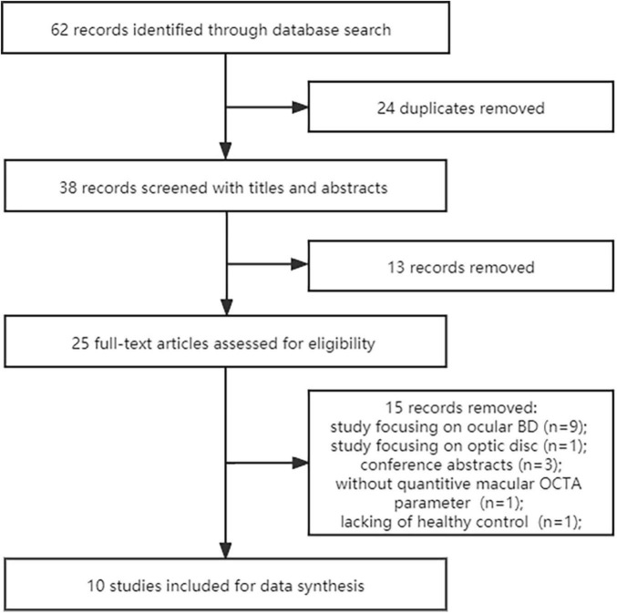
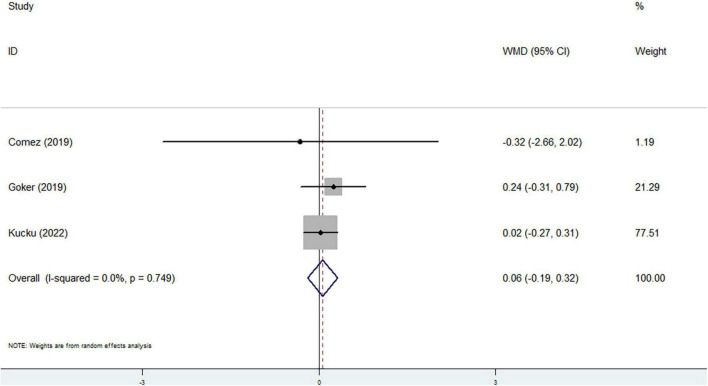
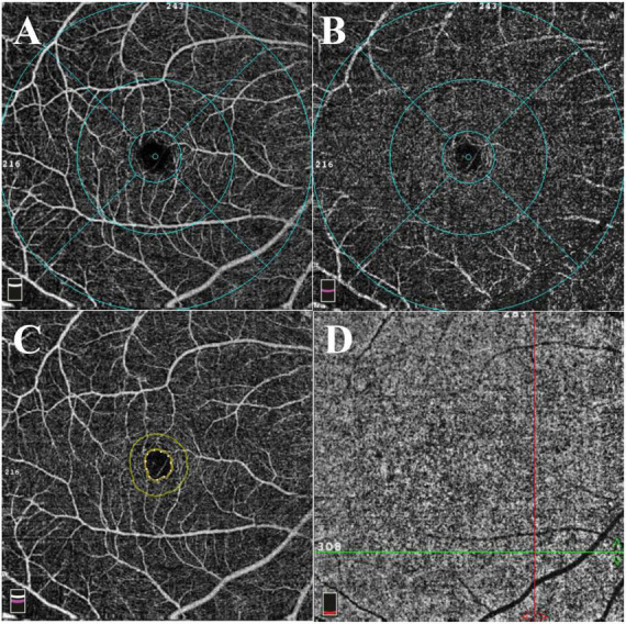
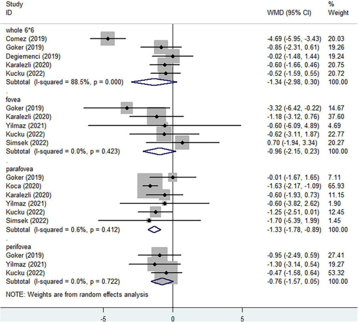
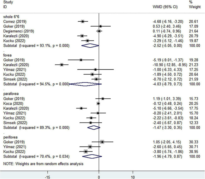
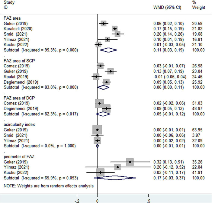
Ten cross-sectional studies involving 386 eyes in non-ocular BD and 418 eyes in healthy volunteers were ultimately included in the present analysis. When considering superficial capillary plexus (SCP) and deep capillary plexus (DCP), no significant differences of vessel densities in the whole enface image, fovea and perifovea were evaluated between two groups. Significantly reduced parafoveal vessel density of SCP was observed in non-ocular BD in comparison with healthy group (WMD = -1.33, 95%CI: -1.78, -0.89; = 0.6%), while slightly decreased parafoveal vessel density was assessed in DCP (WMD = -1.47, 95%CI: -3.30, 0.35; = 89.3%). Significantly increasing foveal avascular zone (FAZ) area was observed in non-ocular BD when compared to healthy controls (WMD = 0.11, 95%CI: 0.03, 0.19; = 95.3%). There was no significant difference in flow area of choriocapillaris between non-ocular BD and control group (WMD = 0.06, 95%CI: -0.19, 0.32; = 0%).

CONCLUSION

Based on current analysis, our results demonstrated significantly lower parafoveal vessel density of SCP and lager FAZ area in full vasculature in non-ocular BD. The retinal microvascular alterations appear before the emergence of ocular manifestations.

SYSTEMATIC TRIAL REGISTRATION

[https://www.crd.york.ac.uk/PROSPERO/], identifier [CRD42021244856].

摘要

目的

我们进行了一项系统评价和荟萃分析,以使用光学相干断层扫描血管造影(OCTA)检查非眼部白塞病(BD)的微血管改变。

方法

在Pubmed、Embase和Cochrane数据库中进行全面检索,以查找从开始到2022年2月的符合条件的研究。由两名独立审阅者从每项研究中提取详细的临床人口统计学数据。加权平均差(WMD)和95%置信区间(CI)用于比较非眼部BD与健康对照之间的OCTA参数。采用Stata 12.0进行统计分析。

结果

本分析最终纳入了10项横断面研究,涉及非眼部BD的386只眼和健康志愿者的418只眼。在考虑浅表毛细血管丛(SCP)和深部毛细血管丛(DCP)时,两组在整个表面图像、黄斑中心凹和黄斑旁区域的血管密度没有显著差异。与健康组相比,非眼部BD患者的SCP黄斑旁血管密度显著降低(WMD = -1.33,95%CI:-1.78,-0.89;P = 0.6%),而DCP的黄斑旁血管密度略有降低(WMD = -1.47,95%CI:-3.30,0.35;P = 89.3%)。与健康对照相比,非眼部BD患者的黄斑无血管区(FAZ)面积显著增加(WMD = 0.11,95%CI:0.03,0.19;P = 95.3%)。非眼部BD与对照组之间的脉络膜毛细血管血流面积没有显著差异(WMD = 0.06,95%CI:-0.19,0.32;P = 0%)。

结论

基于目前的分析,我们的结果表明,非眼部BD患者的SCP黄斑旁血管密度显著降低,全视网膜血管系统的FAZ面积更大。视网膜微血管改变出现在眼部表现出现之前。

系统试验注册

[https://www.crd.york.ac.uk/PROSPERO/],标识符[CRD42021244856]。

https://cdn.ncbi.nlm.nih.gov/pmc/blobs/3429/9353174/ef1eb70413bd/fmed-09-911990-g006.jpg
https://cdn.ncbi.nlm.nih.gov/pmc/blobs/3429/9353174/4034e249c18b/fmed-09-911990-g001.jpg
https://cdn.ncbi.nlm.nih.gov/pmc/blobs/3429/9353174/aa0c5b05c693/fmed-09-911990-g002.jpg
https://cdn.ncbi.nlm.nih.gov/pmc/blobs/3429/9353174/bc22f3d5493b/fmed-09-911990-g003.jpg
https://cdn.ncbi.nlm.nih.gov/pmc/blobs/3429/9353174/2cf714d465ba/fmed-09-911990-g004.jpg
https://cdn.ncbi.nlm.nih.gov/pmc/blobs/3429/9353174/3ea673b24263/fmed-09-911990-g005.jpg
https://cdn.ncbi.nlm.nih.gov/pmc/blobs/3429/9353174/ef1eb70413bd/fmed-09-911990-g006.jpg

相似文献

1
Retinal and choroidal microvascular alterations in Behcet's disease without ocular manifestations: A systematic review and meta-analysis.无眼部表现的白塞病视网膜和脉络膜微血管改变:一项系统评价和荟萃分析。
Front Med (Lausanne). 2022 Jul 22;9:911990. doi: 10.3389/fmed.2022.911990. eCollection 2022.
2
Comparative analysis of optical coherence tomography angiography (OCTA) results between Behçet's disease patients and a healthy control group.比较 Behcet 病患者和健康对照组的光学相干断层扫描血管造影(OCTA)结果。
Clin Rheumatol. 2024 Mar;43(3):1155-1170. doi: 10.1007/s10067-024-06874-y. Epub 2024 Jan 29.
3
Quantitative Analysis of Optical Coherence Tomography Angiography Features in Patients with Nonocular Behcet's Disease.非眼部贝赫切特病患者光学相干断层扫描血管造影特征的定量分析。
Curr Eye Res. 2019 Feb;44(2):212-218. doi: 10.1080/02713683.2018.1530361. Epub 2018 Oct 9.
4
Retinal microvasculature features in patients with Behcet's disease: a systematic review and meta-analysis.贝赫切特病患者的视网膜微血管特征:系统评价和荟萃分析。
Sci Rep. 2022 Jan 14;12(1):752. doi: 10.1038/s41598-021-04730-6.
5
[Correlation of capillary plexus with visual acuity in idiopathic macular epiretinal membrane eyes using optical coherence tomography angiography].[利用光学相干断层扫描血管造影术研究特发性黄斑视网膜前膜眼中毛细血管丛与视力的相关性]
Zhonghua Yan Ke Za Zhi. 2019 Oct 11;55(10):757-762. doi: 10.3760/cma.j.issn.0412-4081.2019.10.006.
6
Vascular Abnormalities in Peripapillary and Macular Regions of Behcet's Uveitis Patients Evaluated by Optical Coherence Tomography Angiography.光学相干断层扫描血管造影评估白塞氏葡萄膜炎患者视乳头周围和黄斑区的血管异常
Front Med (Lausanne). 2021 Sep 16;8:727151. doi: 10.3389/fmed.2021.727151. eCollection 2021.
7
Optical coherence tomography angiography of subclinical ocular features in pediatric Behçet disease.儿童贝赫切特病的亚临床眼部特征的光相干断层扫描血管造影。
J AAPOS. 2022 Feb;26(1):24.e1-24.e6. doi: 10.1016/j.jaapos.2021.10.004. Epub 2022 Jan 12.
8
Quantitative analysis of retinal microcirculation in optical coherence tomography angiography in cases with Behçet's disease without ocular involvement.白塞病非眼部受累病例的光学相干断层扫描血管造影术中视网膜微循环的定量分析。
Int Ophthalmol. 2019 Oct;39(10):2213-2221. doi: 10.1007/s10792-018-1059-z. Epub 2019 Mar 15.
9
Preclinical ocular features in patients with Behçet's disease detected by optical coherence tomography angiography.应用光学相干断层血管造影术检测 Behçet 病患者的临床前眼部特征。
Eye (Lond). 2021 Oct;35(10):2719-2726. doi: 10.1038/s41433-020-01294-z. Epub 2020 Nov 24.
10
Analysis of macular microvasculature with optical coherence tomography angiography for migraine: A systematic review and meta-analysis.利用光学相干断层扫描血管造影术分析偏头痛患者的黄斑微血管:一项系统评价和荟萃分析。
Front Neurol. 2022 Oct 13;13:1001304. doi: 10.3389/fneur.2022.1001304. eCollection 2022.

引用本文的文献

1
Ophthalmic Posterior Segment OCTA Metrics as Potential Biomarkers for Systemic Involvement in Systemic Sclerosis, Systemic Lupus Erythematosus, and Behçet Disease: A Systematic Review.眼科后段光学相干断层扫描血管造影指标作为系统性硬化症、系统性红斑狼疮和白塞病全身受累的潜在生物标志物:一项系统评价
Open Access Rheumatol. 2025 Apr 26;17:87-100. doi: 10.2147/OARRR.S511810. eCollection 2025.

本文引用的文献

1
Vascular complications of Behçet disease.贝切特病的血管并发症。
Minerva Med. 2021 Dec;112(6):767-778. doi: 10.23736/S0026-4806.21.07490-5.
2
Combining retinal and choroidal microvascular metrics improves discriminative power for diabetic retinopathy.视网膜和脉络膜微血管指标的联合应用提高了糖尿病视网膜病变的鉴别能力。
Br J Ophthalmol. 2023 Jul;107(7):993-999. doi: 10.1136/bjophthalmol-2021-319739. Epub 2022 Feb 9.
3
Quantitative changes in peripapillary, macular, and choriocapillaris microvasculature of patients with non-ocular Behçet's disease and relationship with systemic vascular involvement, an optical coherence tomography angiography study.
非眼部贝赫切特病患者的视盘周围、黄斑和脉络膜毛细血管微血管的定量变化及其与系统性血管受累的关系:一项光学相干断层血管造影研究。
Photodiagnosis Photodyn Ther. 2022 Jun;38:102749. doi: 10.1016/j.pdpdt.2022.102749. Epub 2022 Feb 3.
4
Evaluation of retinal and choroidal microcirculation in Behçet's disease.评估贝切特病的视网膜和脉络膜微循环。
Eye (Lond). 2022 Jul;36(7):1494-1499. doi: 10.1038/s41433-022-01932-8. Epub 2022 Feb 4.
5
Retinal microvasculature features in patients with Behcet's disease: a systematic review and meta-analysis.贝赫切特病患者的视网膜微血管特征:系统评价和荟萃分析。
Sci Rep. 2022 Jan 14;12(1):752. doi: 10.1038/s41598-021-04730-6.
6
Parafoveal Microvascular Alterations in Ocular and Non-Ocular Behҫet's Disease Evaluated With Optical Coherence Tomography Angiography.用光学相干断层血管造影术评估眼部和非眼部贝赫切特病的旁中心区微血管改变。
Invest Ophthalmol Vis Sci. 2021 Mar 1;62(3):8. doi: 10.1167/iovs.62.3.8.
7
Optical coherence tomography angiography findings in patients with ocular and non-ocular Behcet disease.眼内和眼外 Behcet 病患者的光学相干断层扫描血管造影表现。
Arq Bras Oftalmol. 2021 May-Jun;84(3):235-240. doi: 10.5935/0004-2749.20210031.
8
Preclinical ocular features in patients with Behçet's disease detected by optical coherence tomography angiography.应用光学相干断层血管造影术检测 Behçet 病患者的临床前眼部特征。
Eye (Lond). 2021 Oct;35(10):2719-2726. doi: 10.1038/s41433-020-01294-z. Epub 2020 Nov 24.
9
Swept-source optical coherence tomography imaging of the retinochoroid and beyond.扫频源光相干断层扫描成像在视网膜脉络膜及其以外的应用。
Expert Rev Med Devices. 2020 May;17(5):413-426. doi: 10.1080/17434440.2020.1755256. Epub 2020 Apr 20.
10
Quantitative Evaluation of the Retinal Vascular Parameters With OCTA in Patients With Behçet Disease Without Ocular Involvement.光学相干断层扫描血管造影对无眼部受累的白塞病患者视网膜血管参数的定量评估
Ophthalmic Surg Lasers Imaging Retina. 2019 Dec 1;51(1):31-34. doi: 10.3928/23258160-20191211-04.