Johnston Noémie, De Rycke Jeffrey, Lievens Yolande, van Eijkeren Marc, Aelterman Jan, Vandersmissen Eva, Ponte Stephan, Vanderstraeten Barbara
Centre Hospitalier Universitaire de Liège, Service de Radiothérapie, Liège, Belgium.
Ghent University, Faculty of Medicine and Health Sciences, Department of Human Structure and Repair, Gent, Belgium.
Phys Imaging Radiat Oncol. 2022 Jul 25;23:109-117. doi: 10.1016/j.phro.2022.07.004. eCollection 2022 Jul.
The geometrical accuracy of auto-segmentation using convolutional neural networks (CNNs) has been demonstrated. This study aimed to investigate the dose-volume impact of differences between automatic and manual OARs for locally advanced (LA) and peripherally located early-stage (ES) non-small cell lung cancer (NSCLC).
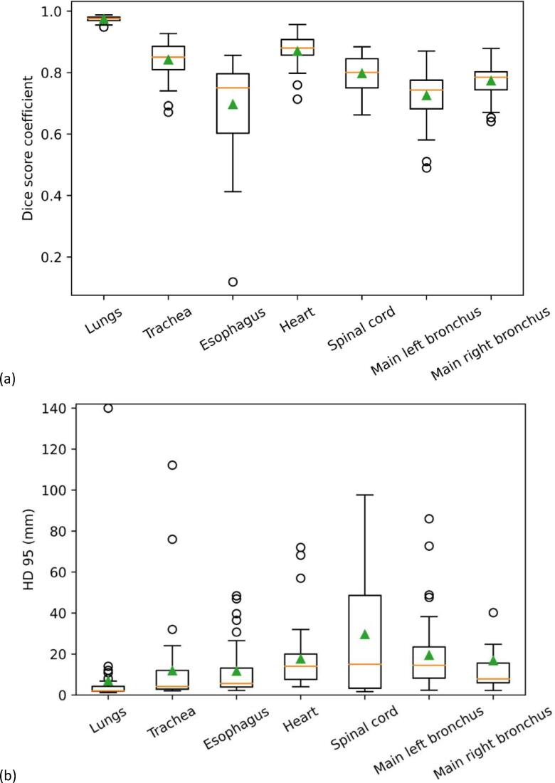
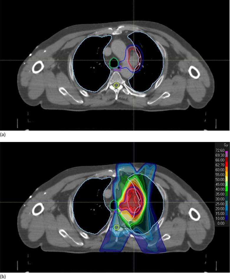
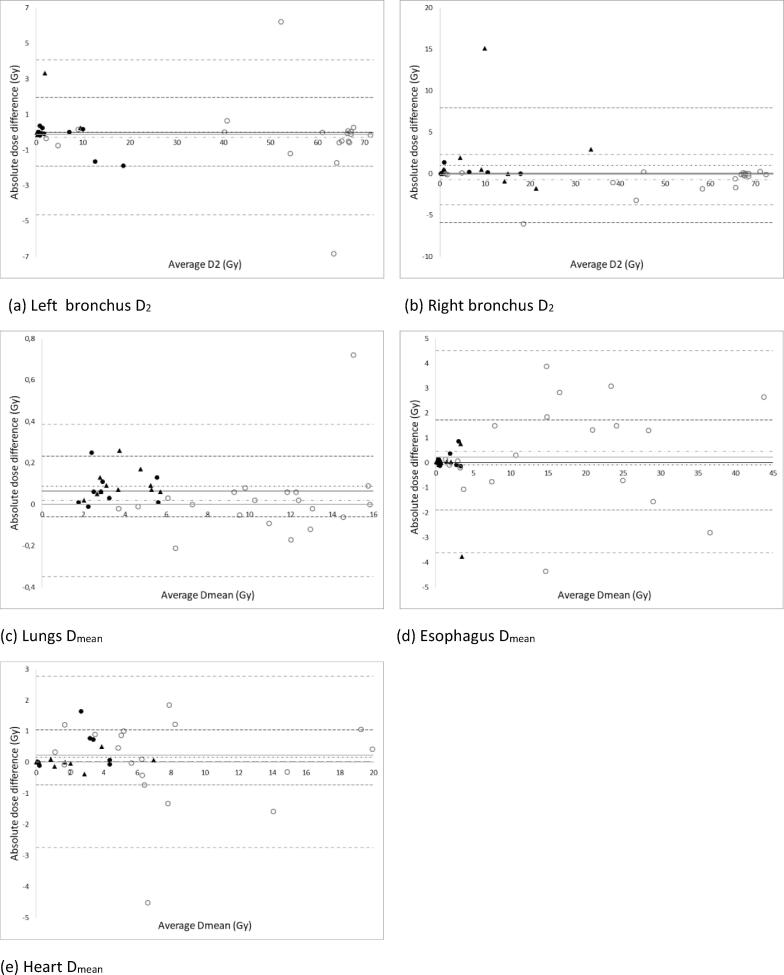
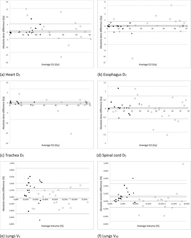
A single CNN was created for automatic delineation of the heart, lungs, main left and right bronchus, esophagus, spinal cord and trachea using 55/10/40 patients for training/validation/testing. Dice score coefficient (DSC) and 95th percentile Hausdorff distance (HD95) were used for geometrical analysis. A new treatment plan based on the auto-segmented OARs was created for each test patient using 3D for ES-NSCLC (SBRT, 3-8 fractions) and IMRT for LA-NSCLC (24-35 fractions). The correlation between geometrical metrics and dose-volume differences was investigated.
The average (±1 SD) DSC and HD95 were 0.82 ± 0.07 and 16.2 ± 22.4 mm, while the average dose-volume differences were 0.5 ± 1.5 Gy (ES) and 1.5 ± 2.8 Gy (LA). The geometrical metrics did not correlate with the observed dose-volume differences (average Pearson for DSC: -0.27 ± 0.18 (ES) and -0.09 ± 0.12 (LA); HD95: 0.1 ± 0.3 mm (ES) and 0.2 ± 0.2 mm (LA)).
After post-processing, manual adjustments of automatic contours are only needed for clinically relevant OARs situated close to the tumor or within an entry or exit beam e.g., the heart and the esophagus for LA-NSCLC and the bronchi for ES-NSCLC. The lungs do not need to be checked further in detail.
已证实使用卷积神经网络(CNN)进行自动分割的几何准确性。本研究旨在调查局部晚期(LA)和周围型早期(ES)非小细胞肺癌(NSCLC)自动和手动勾画的危及器官(OARs)之间差异对剂量体积的影响。
创建了一个单一的CNN,用于自动勾画心脏、肺、左右主支气管、食管、脊髓和气管,使用55/10/40例患者进行训练/验证/测试。使用骰子分数系数(DSC)和第95百分位数豪斯多夫距离(HD95)进行几何分析。对于每个测试患者,基于自动分割的OARs创建了一个新的治疗计划,ES-NSCLC采用三维适形放疗(SBRT,3-8次分割),LA-NSCLC采用调强放疗(IMRT,24-35次分割)。研究了几何指标与剂量体积差异之间的相关性。
平均(±1标准差)DSC和HD95分别为0.82±0.07和16.2±22.4mm,而平均剂量体积差异为0.5±1.5Gy(ES)和1.5±2.8Gy(LA)。几何指标与观察到的剂量体积差异无关(DSC的平均皮尔逊相关系数:-0.27±0.18(ES)和-0.09±0.12(LA);HD95:0.1±0.3mm(ES)和0.2±0.2mm(LA))。
经过后处理后,仅需对位于肿瘤附近或射野入射或出射路径内的临床相关OARs进行手动调整,例如LA-NSCLC的心脏和食管以及ES-NSCLC的支气管。肺部无需进一步详细检查。