Ellistasari Endra Yustin, Kariosentono Harijono, Purwanto Bambang, Wasita Brian, Riswiyant Risya Cilmiaty Arief, Pamungkasari Eti Poncorini, Soetrisno Soetrisno
Doctoral Program of Medical Sciences Department, Sebelas Maret University, Surakarta, Indonesia.
Dermatology and Venereology Department, Sebelas Maret University, Surakarta, Indonesia.
Clin Cosmet Investig Dermatol. 2022 Aug 8;15:1583-1591. doi: 10.2147/CCID.S371330. eCollection 2022.
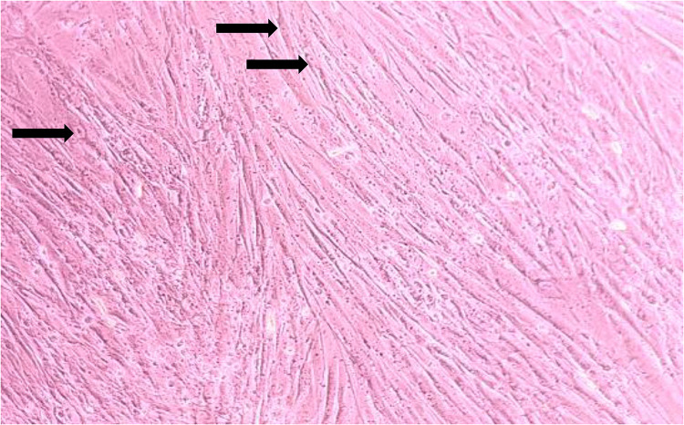
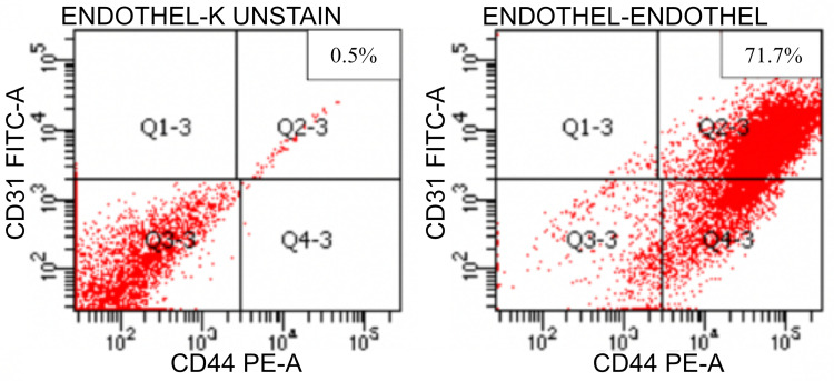
This is an in-vitro experimental study to analyze the effect of Exo-HUVEC on endothelial cell (CD31), cell proliferation, matrix metalloproteinase 1 (MMP-1) and collagen type 1 on irradiated fibroblast with UVB as photo-aging model.
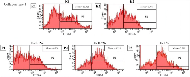
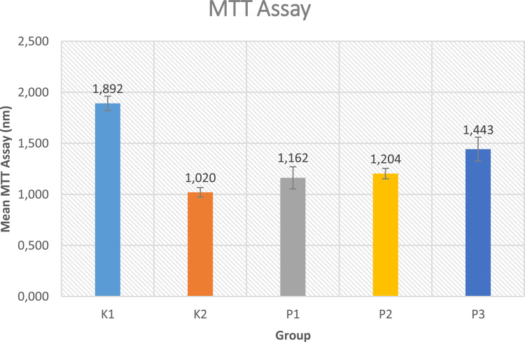
Fibroblast cultures were divided into 5 groups, namely without UVB exposure, UVB exposure 600mJ/cm for 80 seconds as photo-aging model, and UVB exposure +Exo-HUVEC exposure 0.1%, 0.5% and 1%. The endothelial cell was stained with a CD31 marker, MMP-1 were examined with ELISA, cell proliferation is detected using an MTT assay; meanwhile, collagen type 1 deposition and endothelial cell were measured using flowcytometry.
This study found positive endothelial cell marker CD31. Significant difference was found in cell proliferation, MMP-1 and collagen type 1 level between the control group with UVB irradiation and the treatment group with Exo-HUVEC (p < 0.05).
Exo-HUVEC significantly increases cell proliferation and collagen type 1 level, while decrease MMP-1 levels on irradiated fibroblast; therefore, Exo-HUVEC ameliorate the photo-aging of skin fibroblast.
本研究为体外实验研究,以UVB照射成纤维细胞建立光老化模型,分析人脐静脉内皮细胞来源外泌体(Exo-HUVEC)对内皮细胞(CD31)、细胞增殖、基质金属蛋白酶1(MMP-1)和I型胶原蛋白的影响。
将成纤维细胞培养物分为5组,即未暴露于UVB组、以600mJ/cm²照射80秒作为光老化模型的UVB暴露组,以及UVB暴露+0.1%、0.5%和1%的Exo-HUVEC暴露组。用CD31标记物对内皮细胞进行染色,用ELISA检测MMP-1,用MTT法检测细胞增殖;同时,用流式细胞术检测I型胶原蛋白沉积和内皮细胞。
本研究发现内皮细胞标志物CD31呈阳性。在UVB照射的对照组和Exo-HUVEC治疗组之间,细胞增殖、MMP-1和I型胶原蛋白水平存在显著差异(p<0.05)。
Exo-HUVEC可显著提高照射后成纤维细胞的细胞增殖和I型胶原蛋白水平,同时降低MMP-1水平;因此,Exo-HUVEC可改善皮肤成纤维细胞的光老化。