Department of Pediatric Dentistry, Affiliated Stomatology Hospital of Guangzhou Medical University, Guangdong Engineering Research Center of Oral Restoration and Reconstruction, Guangzhou Key Laboratory of Basic and Applied Research of Oral Regenerative Medicine, Guangzhou, China.
J Cell Mol Med. 2023 Apr;27(8):1131-1143. doi: 10.1111/jcmm.17727. Epub 2023 Mar 25.
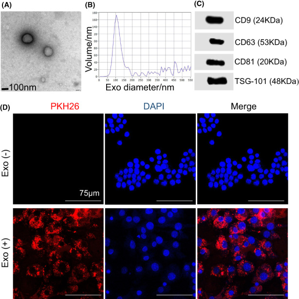
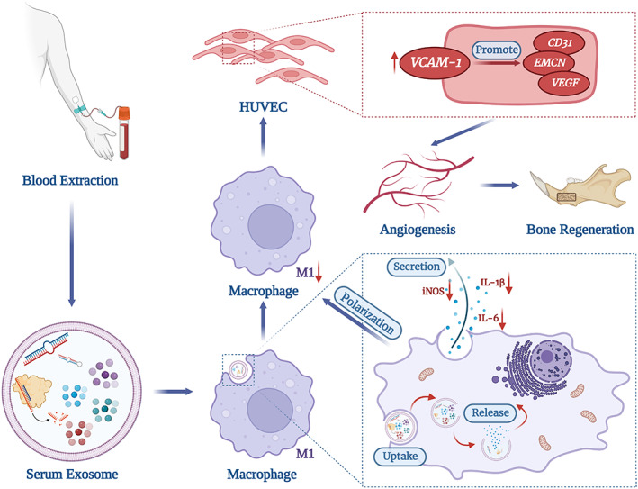
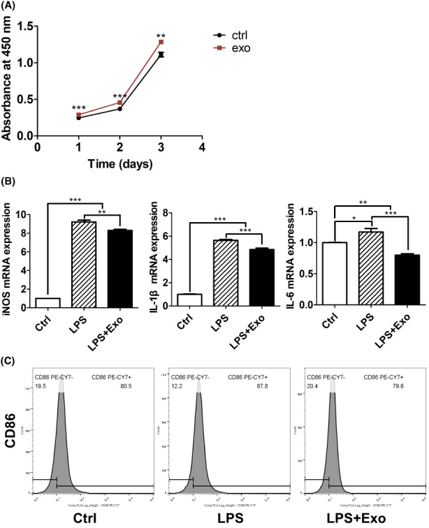
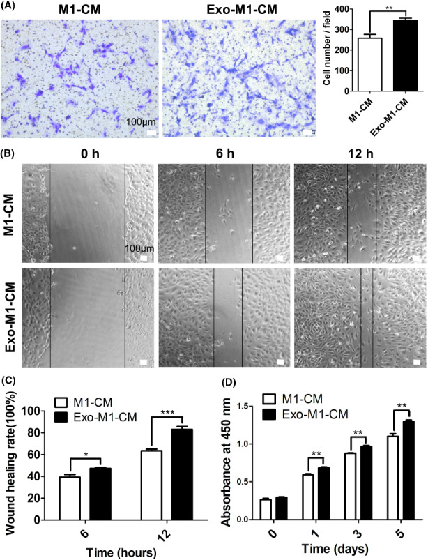
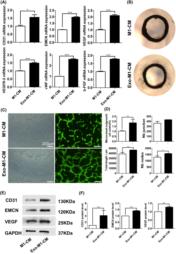
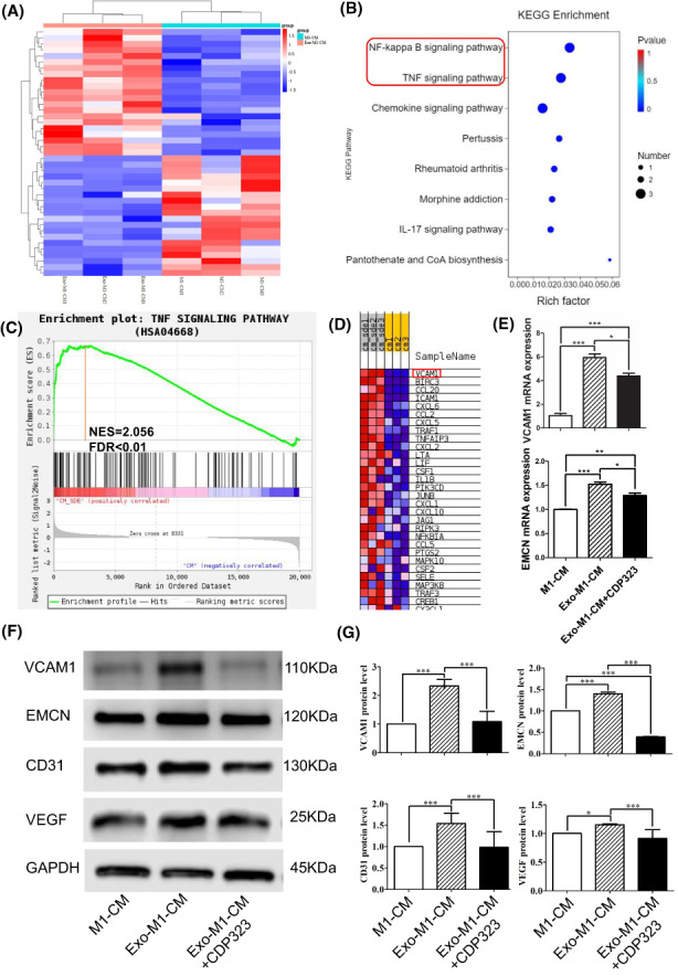
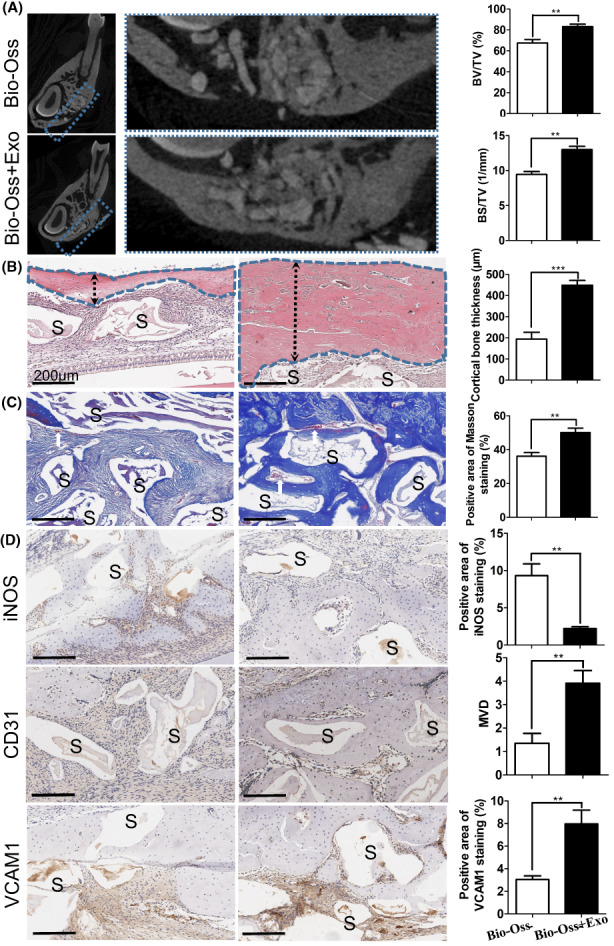
During exogenous bone-graft-mediated bone defect repair, macrophage inflammation dictates angiogenesis and bone regeneration. Exosomes from different human cells have shown macrophage immunomodulation-mediated bone regeneration potential. However, the effect of human serum-derived exosomes (serum-Exo) on macrophage immunomodulation-mediated angiogenesis during bone defect repair has not been investigated yet. In this study, we explored the effects of serum-Exo on macrophage inflammation regulation-mediated angiogenesis during bone defect repair and preliminarily elucidated the mechanism. Healthy serum-Exo was isolated by ultracentrifugation. The effect of serum-Exo on LPS-induced M1 macrophage inflammation was analysed in vitro. The conditioned medium of serum-Exo-treated LPS-induced M1 macrophage (serum-Exo-treated M1 macrophage-CM) was used to culture human umbilical vein endothelial cells (HUVEC), and the effect on angiogenesis was analysed by western blot, qRT-PCR, etc. mRNA-sequencing of HUVECs was performed to identify deferentially expressed genes. Finally, the rat mandibular defect model was established and treated with Bio-Oss and Bio-Oss + Exo. The effect of the Bio-Oss + Exo combination on mandibular bone regeneration was observed by micro-computed tomography (micro-CT), haematoxylin and eosin (HE) staining, Masson staining, and immunohistochemical staining. Serum-Exo promoted the proliferation of RAW264.7 macrophages and reduced the expression of M1-related genes such as IL-6, IL-1β, iNOS, and CD86. Serum-Exo-treated M1 macrophage-CM induced the proliferation, migration, and angiogenic differentiation of HUVEC, as well as the expression of H-type blood vessel markers CD31 and endomucin (EMCN), compared with M1 macrophage-CM. Moreover, higher expression of vascular endothelial adhesion factor 1 (VCAM1) in HUVEC cultured with serum-Exo-treated M1 macrophage-CM compared with M1 macrophages-CM. Inhibition of VCAM1 signalling abrogated the pro-angiogenic effect of serum-Exo-treated M1 macrophage-CM on HUVEC. Local administration of serum-Exo during mandibular bone defect repair reduced the number of M1 macrophages and promoted angiogenesis and osteogenesis. Collectively, our results demonstrate the macrophage inflammation regulation-mediated pro-angiogenic potential of serum-Exo during bone defect repair possibly via upregulation of VCAM1 signalling in HUVEC.
在骨缺损修复过程中,外源性骨移植介导的骨修复过程中,巨噬细胞炎症决定了血管生成和骨再生。不同来源的细胞外囊泡已显示出通过调节巨噬细胞免疫来促进骨再生的潜力。然而,尚未研究人血清来源的细胞外囊泡(血清外泌体)对骨缺损修复过程中巨噬细胞炎症调节介导的血管生成的影响。在这项研究中,我们探讨了血清外泌体对骨缺损修复过程中巨噬细胞炎症调节介导的血管生成的影响,并初步阐明了其机制。通过超速离心分离健康的血清外泌体。在体外分析血清外泌体对脂多糖诱导的 M1 巨噬细胞炎症的影响。用血清外泌体处理脂多糖诱导的 M1 巨噬细胞(血清外泌体处理的 M1 巨噬细胞-CM)的条件培养基培养人脐静脉内皮细胞(HUVEC),通过 Western blot、qRT-PCR 等方法分析对血管生成的影响。对 HUVEC 进行 mRNA 测序,以鉴定差异表达的基因。最后,建立大鼠下颌骨缺损模型,并分别用 Bio-Oss 和 Bio-Oss+Exo 处理。通过 micro-CT、苏木精和伊红(HE)染色、Masson 染色和免疫组织化学染色观察 Bio-Oss+Exo 组合对下颌骨再生的影响。血清外泌体促进 RAW264.7 巨噬细胞的增殖,并降低 IL-6、IL-1β、iNOS 和 CD86 等 M1 相关基因的表达。与 M1 巨噬细胞-CM 相比,血清外泌体处理的 M1 巨噬细胞-CM 诱导 HUVEC 的增殖、迁移和血管生成分化,并上调 H 型血管标志物 CD31 和内皮黏蛋白(EMCN)的表达。此外,与 M1 巨噬细胞-CM 相比,培养有血清外泌体处理的 M1 巨噬细胞-CM 的 HUVEC 中血管内皮细胞黏附因子 1(VCAM1)的表达更高。抑制 VCAM1 信号通路可消除血清外泌体处理的 M1 巨噬细胞-CM 对 HUVEC 的促血管生成作用。在下颌骨骨缺损修复过程中局部给予血清外泌体可减少 M1 巨噬细胞的数量,并促进血管生成和成骨。总之,我们的研究结果表明,血清外泌体在骨缺损修复过程中通过上调 HUVEC 中的 VCAM1 信号转导来调节巨噬细胞炎症,从而发挥促血管生成作用。