Caltech Optical Imaging Laboratory, Andrew and Peggy Cherng Department of Medical Engineering, California Institute of Technology, Pasadena, CA, 91125, USA.
Lilly Research Laboratories, Eli Lilly and Company, Lilly Corporate Center, Indianapolis, IN, 46285, USA.
Adv Sci (Weinh). 2022 Oct;9(28):e2202907. doi: 10.1002/advs.202202907. Epub 2022 Aug 17.
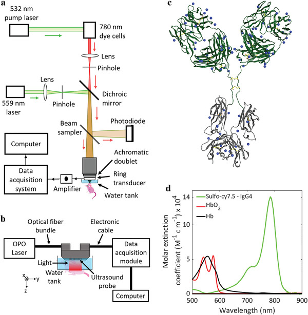
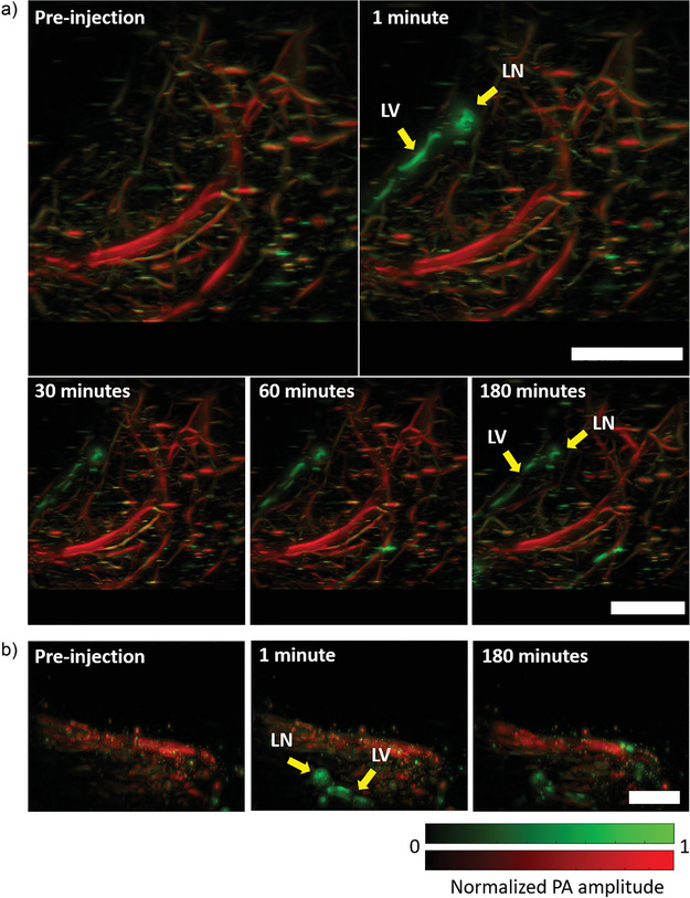
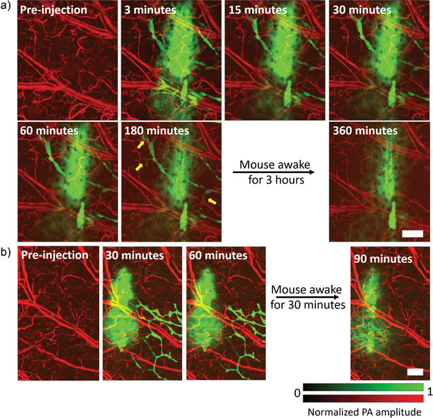
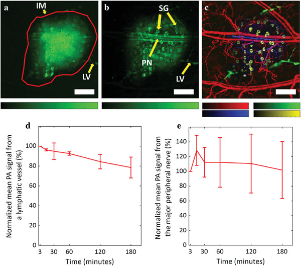
Long-duration in vivo simultaneous imaging of multiple anatomical structures is useful for understanding physiological aspects of diseases, informative for molecular optimization in preclinical models, and has potential applications in surgical settings to improve clinical outcomes. Previous studies involving simultaneous imaging of multiple anatomical structures, for example, blood and lymphatic vessels as well as peripheral nerves and sebaceous glands, have used genetically engineered mice, which require expensive and time-consuming methods. Here, an IgG4 isotype control antibody is labeled with a near-infrared dye and injected into a mouse ear to enable simultaneous visualization of blood and lymphatic vessels, peripheral nerves, and sebaceous glands for up to 3 h using photoacoustic microscopy. For multiple anatomical structure imaging, peripheral nerves and sebaceous glands are imaged inside the injected dye-labeled antibody mass while the lymphatic vessels are visualized outside the mass. The efficacy of the contrast agent to label and localize deep medial lymphatic vessels and lymph nodes using photoacoustic computed tomography is demonstrated. The capability of a single injectable contrast agent to image multiple structures for several hours will potentially improve preclinical therapeutic optimization, shorten discovery timelines, and enable clinical treatments.
长时间在体同时对多个解剖结构进行成像,有助于了解疾病的生理方面,为临床前模型中的分子优化提供信息,并有可能在手术环境中改善临床结果。以前的研究涉及同时对多个解剖结构进行成像,例如血液和淋巴管以及周围神经和皮脂腺,使用了需要昂贵且耗时的方法的基因工程小鼠。在这里,一种 IgG4 同种型对照抗体用近红外染料标记,并注入小鼠耳朵中,使用光声显微镜可在长达 3 小时内同时可视化血液和淋巴管、周围神经和皮脂腺。对于多个解剖结构成像,在注入的染料标记抗体团块内部成像周围神经和皮脂腺,而在团块外部可视化淋巴管。使用光声计算机断层扫描证明了该造影剂标记和定位深部内侧淋巴管和淋巴结的效果。单一可注射造影剂能够在数小时内对多个结构进行成像,这将有可能改善临床前治疗优化、缩短发现时间线并实现临床治疗。